XB-IMG-135226
Xenbase Image ID: 135226
|
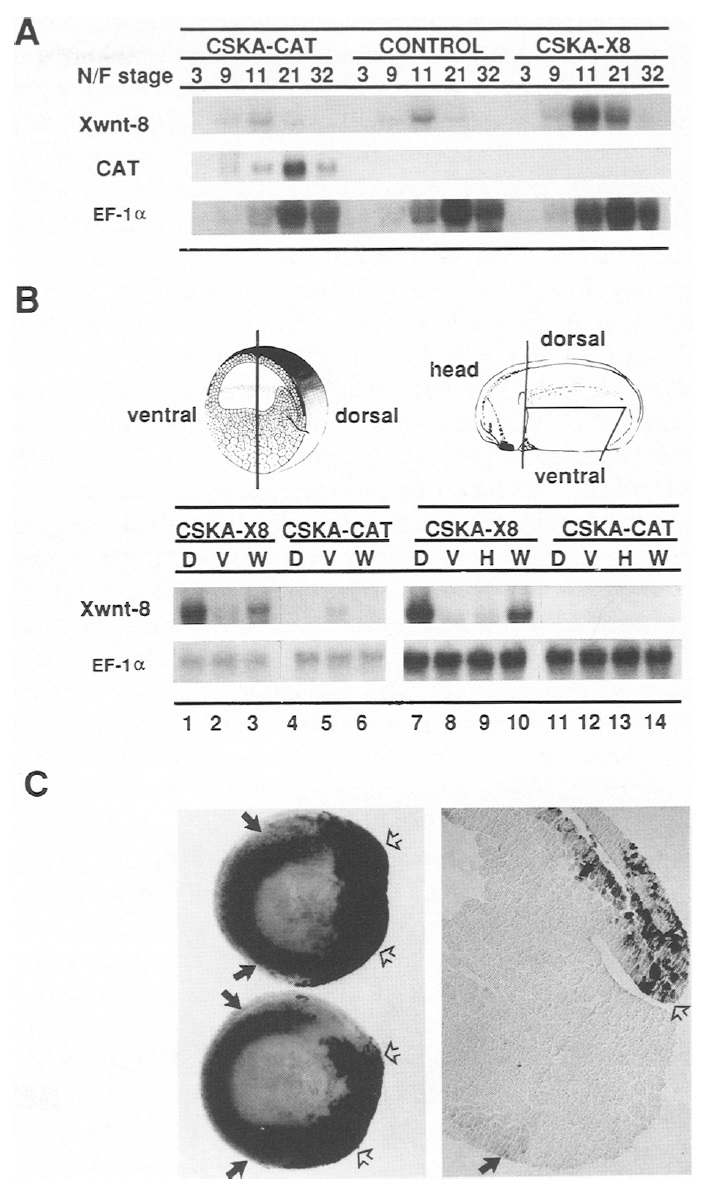
Figure 3. Expression of Xwnt-8 and CAT transcripts following
injection of CSKA-X8 or CSKA-CAT DNA into Xenopus embryos.
(A) RNA was isolated from injected or uninjected embryos
at cleavage (stage 3), blastula (stage 9), gastrula (stage 11 ),
neurula (stage 21), or tailbud (stage 32) stages, as indicated at the
top of A [(N/F) Nieuwkoop and Faber 1967] and subjected to gel
blot analysis. The filter was successively hybridized with the
probes indicated at left. (B) Gastrulae (stage 10; lanes 1-6) or
neurulae (stage 21; lanes 7-14) were dissected as illustrated.
Northern blots containing RNA from the region indicated
above each lane were successively hybridized with the probes
indicated at left. The filter was exposed to film for 10 hr, or for
6 hr, to obtain the Xwnt-8 signals shown for lanes 7-14, and
1-6, respectively. (D)Dorsal; (V) ventral; (W) whole; (H) head.
(C) Whole-mount in situ hybridization of Xwnt-8 probes to
stage-11 gastrulae that had received dorsal injections of CSKAX8.
Solid arrows denote signal from endogenous Xwnt-8 transcripts
in ventrolateral maginal zone ceils; open arrows indicate
staining of dorsal lip cells from plasmid-derived Xwnt-8. (A)
Whole gastrulae; (B) sagittal section through a representative
embryo. Image published in: Christian JL and Moon RT (1993) Copyright © 1993. Image reproduced on with permission of the Publisher, Cold Spring Harbor Laboratory Press. This is an Open Access article distributed under the terms of the Creative Commons Attribution License. Larger Image Printer Friendly View |