XB-IMG-138872
Xenbase Image ID: 138872
|
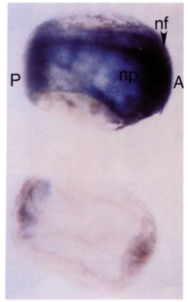
FIG. 7. Localization of XLPOU 1 by whole mount in situ hybridization.
Whole, fixed embryos were hybridized to either sense or antisense
digoxigenin-labeled riboprobes of XLPOU 1 OP at stage 15 (control
is below). The dark blue reaction product representing hybridization
can easily be distinguished from the dark brown pigment found in
wild-type embryos. Embryos hybridized to the sense probe (controls)
were lightly stained in comparison to embryos hybridized to the antisense
probe. The anterior (A) and posterior (P) ends of the embryo,
neural plate (np), and neural folds (nf) are indicated. Image published in: Agarwal VR and Sato SM (1991) Copyright © 1991. Image reproduced with permission of the Publisher, Elsevier B. V.
Image source: Published Larger Image Printer Friendly View |