XB-IMG-145864
Xenbase Image ID: 145864
|
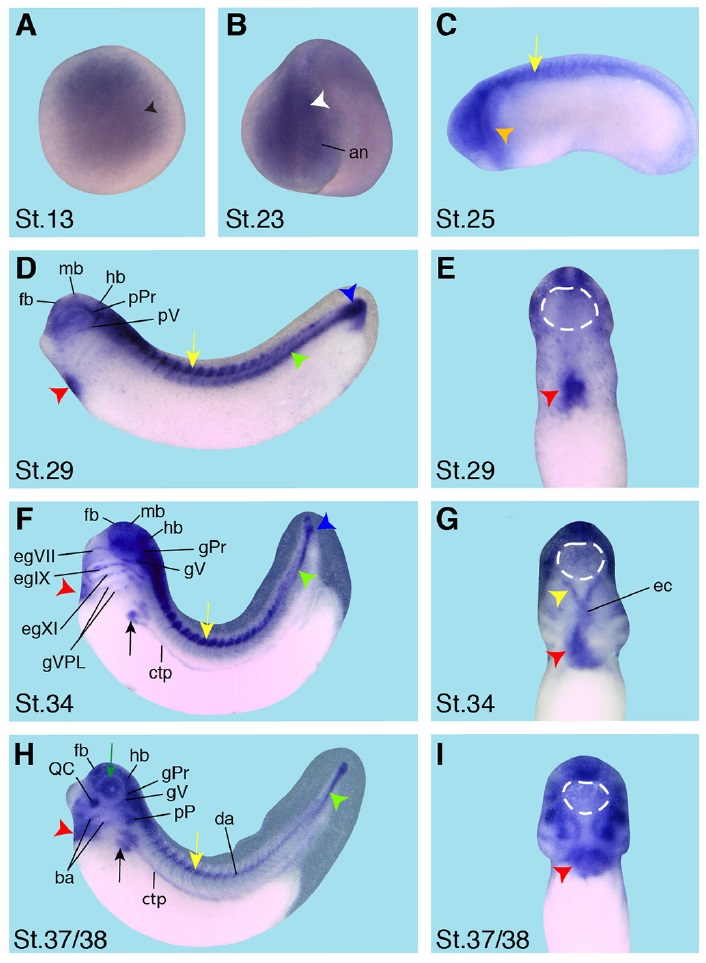
Fig. 6 (Left). Spatial expression pattern of crip2 during X. laevis embryonic development. Embryonic stages are indicated in each panel. The cement gland is depicted as white dashed circles. (A) Animal view of a stage 13 embryo. Crip2 expression was detected in the anterior neural plate (black arrowhead). (B) The anterior view of a stage 23 embryo showed a crip2 expression in the anterior neural tissue (an) and the neural tube (white arrowhead). (C-D,F,H) Lateral views with anterior to the left. (E,G,I) Ventral views with anterior to the top. (C) At stage 25 crip2 transcripts were detected in the migrating cranial neural crest cells (orange arrowhead) and the dorsal side of the embryo (yellow arrow). (D-I) Crip2 was expressed in the cardiac tissue especially of the first heart field (red arrowheads), the endocardium (ec) and the cardiac vascular nerves (yellow arrowhead). (D) At stage 29 crip2 transcripts were strongly detected at the dorsal side (yellow arrow), the posterior cardinal vein (green arrowhead) and at the tip of the tail (blue arrowhead). Furthermore, crip2 was expressed in the fore-, mid- and hindbrain (fb, mb, hb) as well as in the profundal placode (pPr) and the trigeminal placode (pV). (F,H) Later during organogenesis crip2 transcripts were additionally detected in the anterior part of pronephros (black arrow) and the connecting tubule of the pronephros (ctp). (F) At stage 34 the profundal ganglion (gPr), the trigeminal ganglion (gV), the facial epibranchial ganglia egVII, egIX and egXI and the cells that contribute to the vagal and posterior lateral line ganglion (gVPL) were positive for crip2. (H) At stage 37/38 crip2 was also expressed the quadrate cartilages (QC), branchial arches (ba) and dorsal aorta (da). This image is extracted from figure extracted from: Xenbase Image, This image is extracted from figure published in: Hempel A and Kühl SJ (2014), Image published in: Hempel A and Kühl SJ (2014) Copyright © 2014. Reproduced with permission of the Publisher, University of the Basque Country Press.
Image source: Published Larger Image Printer Friendly View |