XB-IMG-172241
Xenbase Image ID: 172241
|
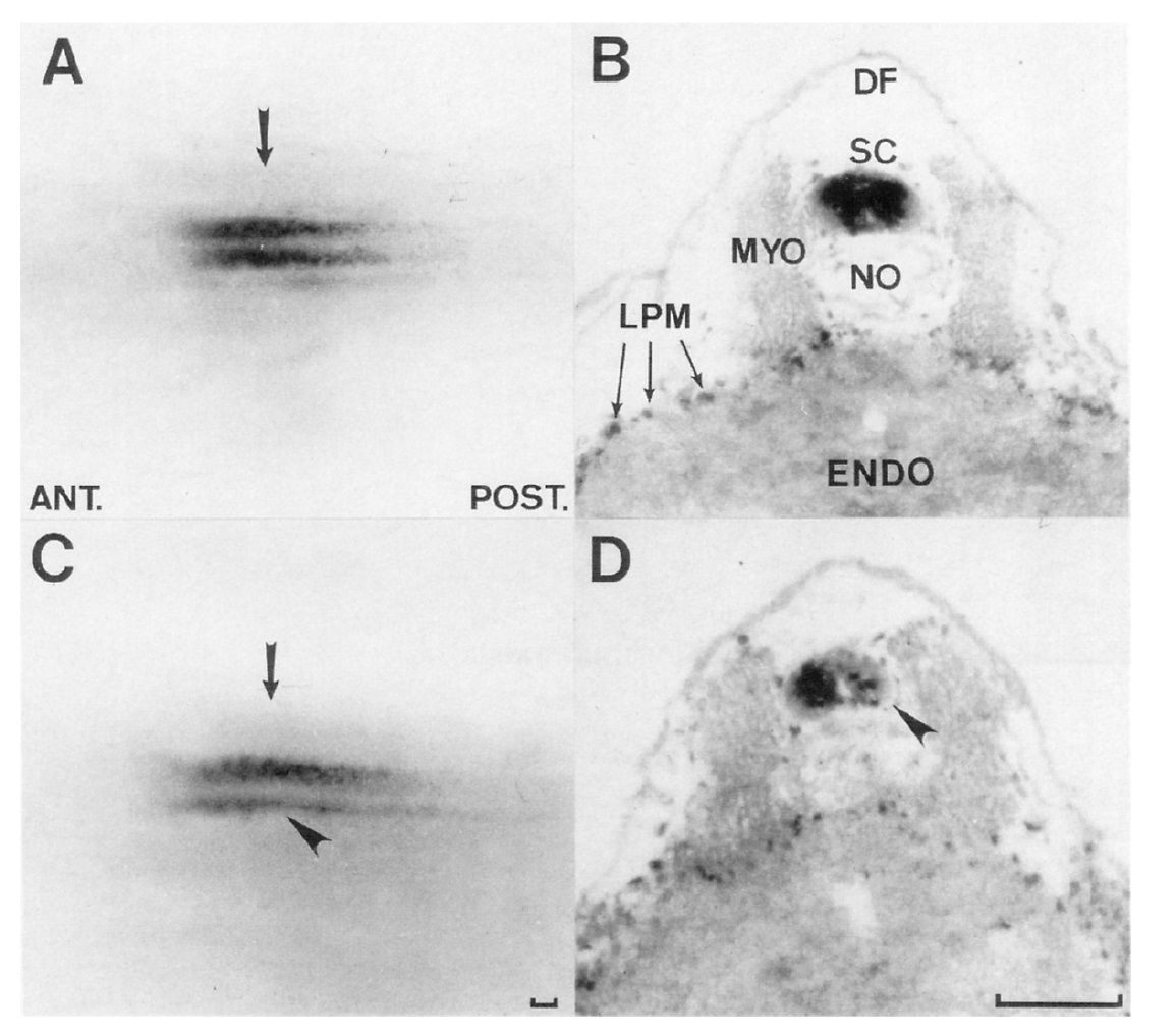
Figure 5. Effect of Unilateral Injection at the
Z-Cell Stage of mRNA Encoding the Short
XlHbox 1 Protein
The top two photographs (A, B) are of an embryo
injected with mutant short XlHbox l/pro45
protein mRNA. Uninjected control tadpoles were
identical. The bottom pair (C, D) corresponds to
an embryo injected with short XlHbox 1 protein
mANA. (A) and (C) show dorsal views of embryos
immunostained in whole mount with long
XlHbox 1Ab. (B) and (D) show transverse sections
taken at the level of the arrows in (A) and
(C), respectively, immunostained with long
XlHbox I-Ab; positive nuclei appear black. Arrowheads
in (C) and (D) indicate the reduced
number of neurons expressing XlHbox 1 caused
by producing short XlHbox 1 protein on this
side of the embryo. In (8) and (D), XlHbox
l-positive nuclei are present in the lateral plate
mesoderm (Oliver et al., 1988; Wright et al.,
1989a). Mydome cells do not express the XlHbox
1 protein. Abbreviations: ANT., anterior; POST.,
posterior; DF, dorsal fin; ENDO, endoderm;
LPM, lateral plate mesoderm; MYO, myotome;
NO, notochord; SC, spinal cord. Bar = 50 pm. Image published in: Wright CV et al. (1989) Copyright © 1989. Image reproduced with permission of the Publisher, Elsevier B. V. Larger Image Printer Friendly View |