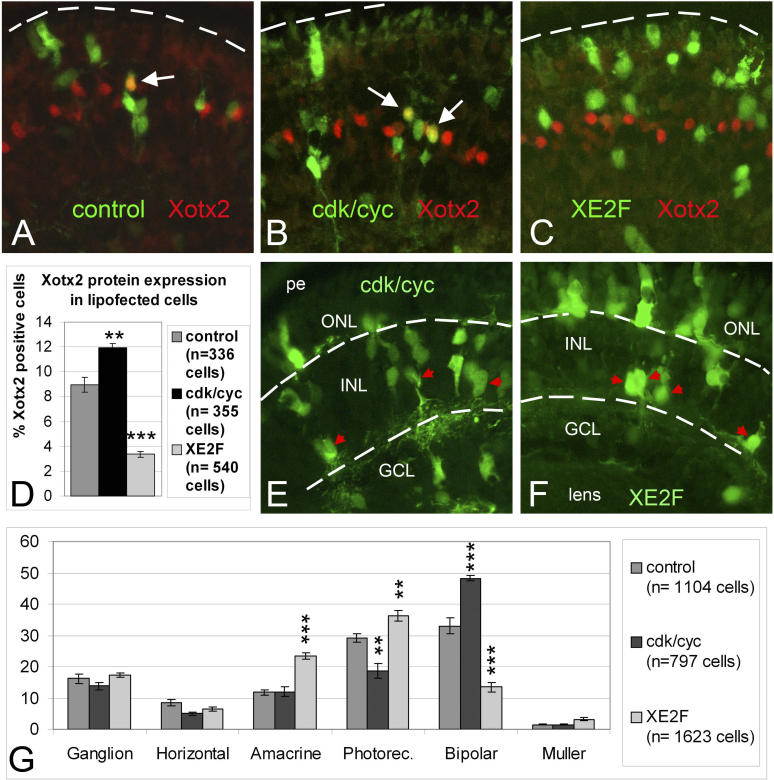
Figure 8. XE2F Lipofection Inhibits Xotx2 Translation and the Generation of Bipolar Cells(AâC) st. 40-lipofected retinas. Lipofected cells are traced by GFP (green), Xotx2 immunostaining in red. Arrows indicate Xotx2-positive lipofected cells.(D) Histogram showing the proportion of retinal lipofected cells expressing Xotx2 protein. Number of counted cells is indicated by n. Double asterisk indicates p = 0.009; triple asterisk indicates p = 0.0001 (student's t-test); error bars indicate standard error of the mean.(EâG) Cell type analysis of lipofected retinas at st. 42. Examples are shown of retinas lipofected with cdk2/cyclinA2 (cdk/cyc in [E]) and XE2F (F). GCL: ganglion cell layer; INL: inner nuclear layer; ONL: outer nuclear layer; pe, pigmented epithelium. Red arrowheads point at cells with amacrine morphology, which represent the majority of cell types in the INL of XE2F-lipofected retinas. Bars in (G) show the proportion of lipofected cells types at st. 42. Number of counted cells is indicated by n; double asterisk indicates p ⤠0.01; triple asterisk indicates p ⤠0.001 (student's t-test); error bars: standard error of the mean. The lipofection of cdk2/cyclinA2 and XE2F increases and decreases, respectively, the proportion of bipolar cells compared to control. The decrease of photoreceptors after cdk2/cyclinA2 lipofection is due to a reduction of cones [18].
Image published in: Decembrini S et al. (2006)
Copyright: © 2006 Decembrini et al. Creative Commons Attribution license
| Gene | Synonyms | Species | Stage(s) | Tissue |
|---|---|---|---|---|
| otx2.L | otx-2, otx2-a, otx2-b, otxA, Xotx-2, Xotx2 | X. laevis | Throughout NF stage 40 | retina bipolar neuron |
Image source: Published
Permanent Image Page
Printer Friendly View
XB-IMG-116832