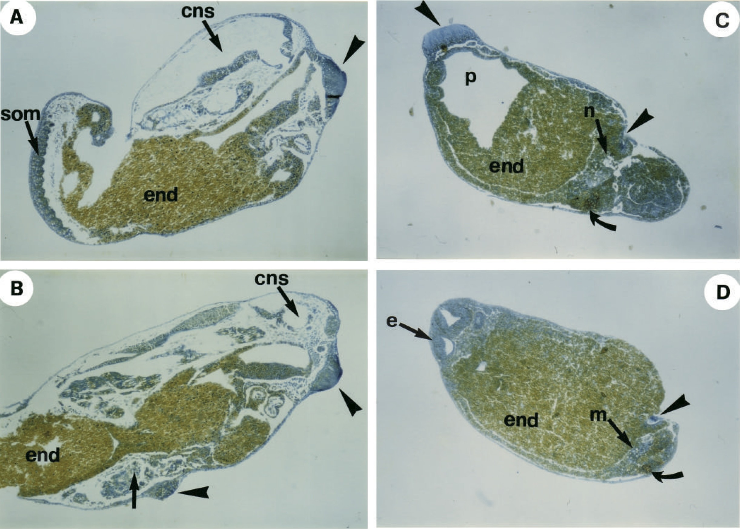
Fig. 8. Histological analysis of embryos microinjected with Xotx2. (A,B) Sagittal sections of embryos of the type in Fig. 7C and D, respectively at a stage corresponding to 34 in control embryos. Arrowheads point to cement glands, whereas an arrow points to ectopic structures located in the vicinity of the secondary cement gland. (C,D) Frontal sections at different levels of embryos of the type in Fig. 7D at a stage corresponding to 32 in control embryos. Fixed embryos were first whole-mount stained with monoclonal antibody 2G9, revealed with an HRP-conjugated secondary antibody and later processed for standard histology. Strong 2G9 staining is detectable among ectopic derivatives (arrows) where, in addition to a few mucus-secreting cement gland cells (arrowhead), muscles (m) and notochord (n) are visible. (cns), central nervous system; (e), eye; (end), endoderm; (p), pharynx; (som), somites.
Image published in: Pannese M et al. (1995)
Copyright © 1995. Image reproduced with permission of the publisher and the copyright holder. This is an Open Access article distributed under the terms of the Creative Commons Attribution License.
Permanent Image Page
Printer Friendly View
XB-IMG-129918