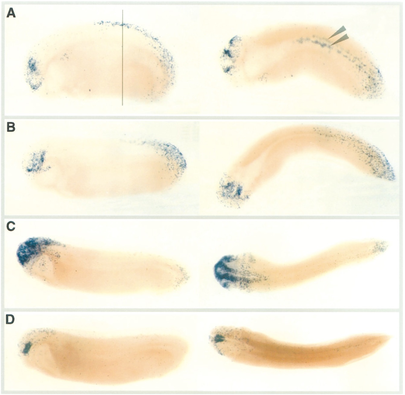
FIG. 5. Programmed cell death as detected by whole-mount TUNEL staining during stages 26–28. Each panel shows a lateral view (left), and a dorsal view (right). (A) Stage 26, the embryo looks younger in the lateral view due to curvature. TUNEL staining is evident in the ventral forebrain and midbrain in addition to the eye vesicle. Staining was also evident in the posterior half of the spinal cord (arrowheads), and in the tailbud. Line indicates the approximate location of the transverse section shown in figure 7D. (B) Stage 26, staining in the midbrain, posterior spinal cord and tailbud. (C) Stages 27–28, staining in the midbrain, hindbrain, and eye vesicles. (D) Stages 27–28, bilaterally symmetrical staining in the midbrain. The patterns of cell death shown are representative of the cell death detected following staining of 36 embryos, all of which were TUNEL positive. Embryos with more than 5 TUNEL-stained nuclei were considered TUNEL positive.
Image published in: Hensey C and Gautier J (1998)
Copyright © 1998. Image reproduced with permission of the Publisher, Elsevier B. V.
Permanent Image Page
Printer Friendly View
XB-IMG-134396