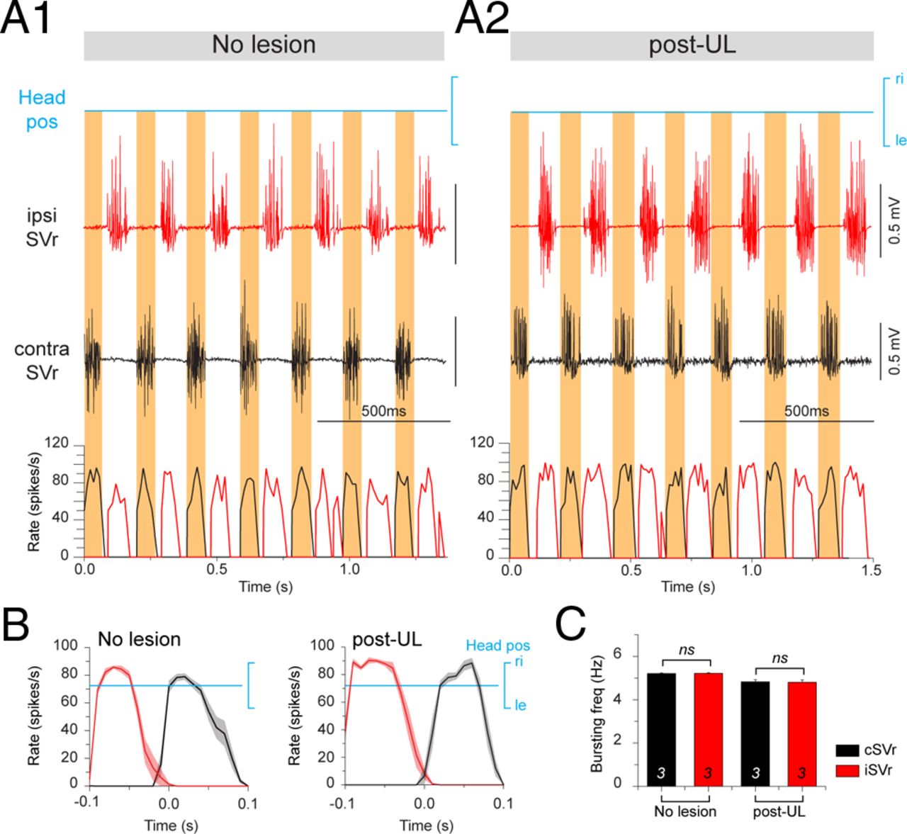
Figure 5. SVr discharge during a spontaneous fictive swimming sequence in stage 56 larval Xenopus before and after UL. A, Spike discharge and firing rate modulation of the SVr on the ipsilesional side (red traces) and contralesional side (black traces) during fictive swimming in a control (A1, no lesion) and 6 weeks post-UL (A2). B, Mean discharge rate of swimming-related bursts over one swim cycle (n = 20; ±SE; shaded area in each plot) of the ipsilesional (red traces) and contralesional (black traces) SVr in a control and after UL. C, Average (±SE) burst frequency of ispilesional and contralesional SVr nerves before (n = 3) and after (n = 3) UL revealed no difference between the two sides, respectively (Wilcoxon signed-rank test for paired parameters; n.s.). ipsi indicates ipsilesional side; contra, contralesional side; ri, right, le, left; i/cSVr, ipsi/contra SVr.
Image published in: Lambert FM et al. (2013)
Copyright © 2013. OA ARTICLE, images redisplayed under a Creative Commons license.
Permanent Image Page
Printer Friendly View
XB-IMG-138219