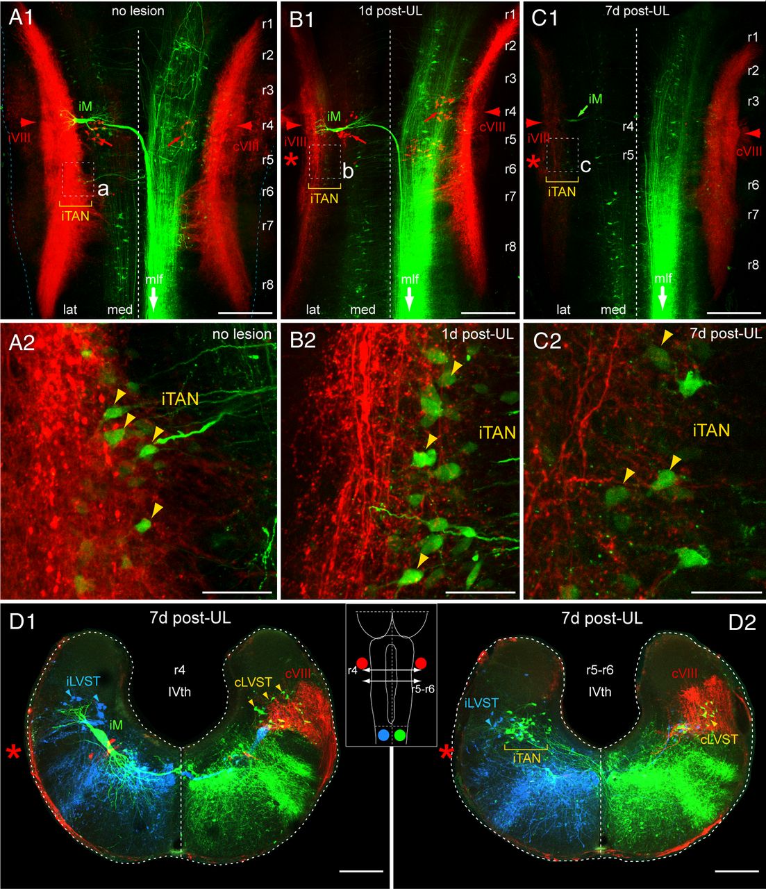
Figure 7. Postlesional loss of vestibular nerve afferent terminations in the vestibular nuclei after UL. AâC, Confocal reconstructions of hindbrain whole-mount preparations of stage 55â56 Xenopus tadpoles showing labeled vestibular afferent terminations on both sides (red, Alexa Fluor 546 dextran) along with contralateral-projecting vestibulo and reticulospinal neurons (green, Alexa Fluor 488 dextran) in controls (A, no lesion), 1 d (B), and 7 d (C) post-UL; higher magnification of the ipsilesional TAN (iTAN; outlined areas in A1âC1) that forms a major subgroup of vestibulospinal neurons (yellow arrowheads), illustrating the successive loss of afferent fibers and terminations on the operated side after UL (B2,C2) with respect to controls (A2). D, Confocal reconstruction of cross-sections of the hindbrain at r4 (D1) and r5âr6 (D2) 7 d after UL on the left side (red asterisks) depicting the location of the Mauthner neuron (M), vestibulospinal neurons that descend in the lateral vestibulospinal tract (LVST), and vestibulospinal neurons in the TAN along with labeled vestibular afferent fibers (red VIII); the schematic inset illustrates the rostrocaudal hindbrain level of the cross-sections, the respective sites of tracer application on the left (blue, Alexa Fluor 647 dextran) and on the right side (green, Alexa Fluor 488 dextran) of the upper spinal cord, and tracer application to the bilateral VIIIth nerve (red, Alexa Fluor 546 dextran). Scale bar in A1âC1 represents 250 μm, in A2âC2 50 μm and in D1,2 200 μm. IVth, IVth ventricle; i, ipsilesional side; c, contralesional side; r1â8, rhombomere 1â8.
Image published in: Lambert FM et al. (2013)
Copyright © 2013. OA ARTICLE, images redisplayed under a Creative Commons license.
Permanent Image Page
Printer Friendly View
XB-IMG-138221