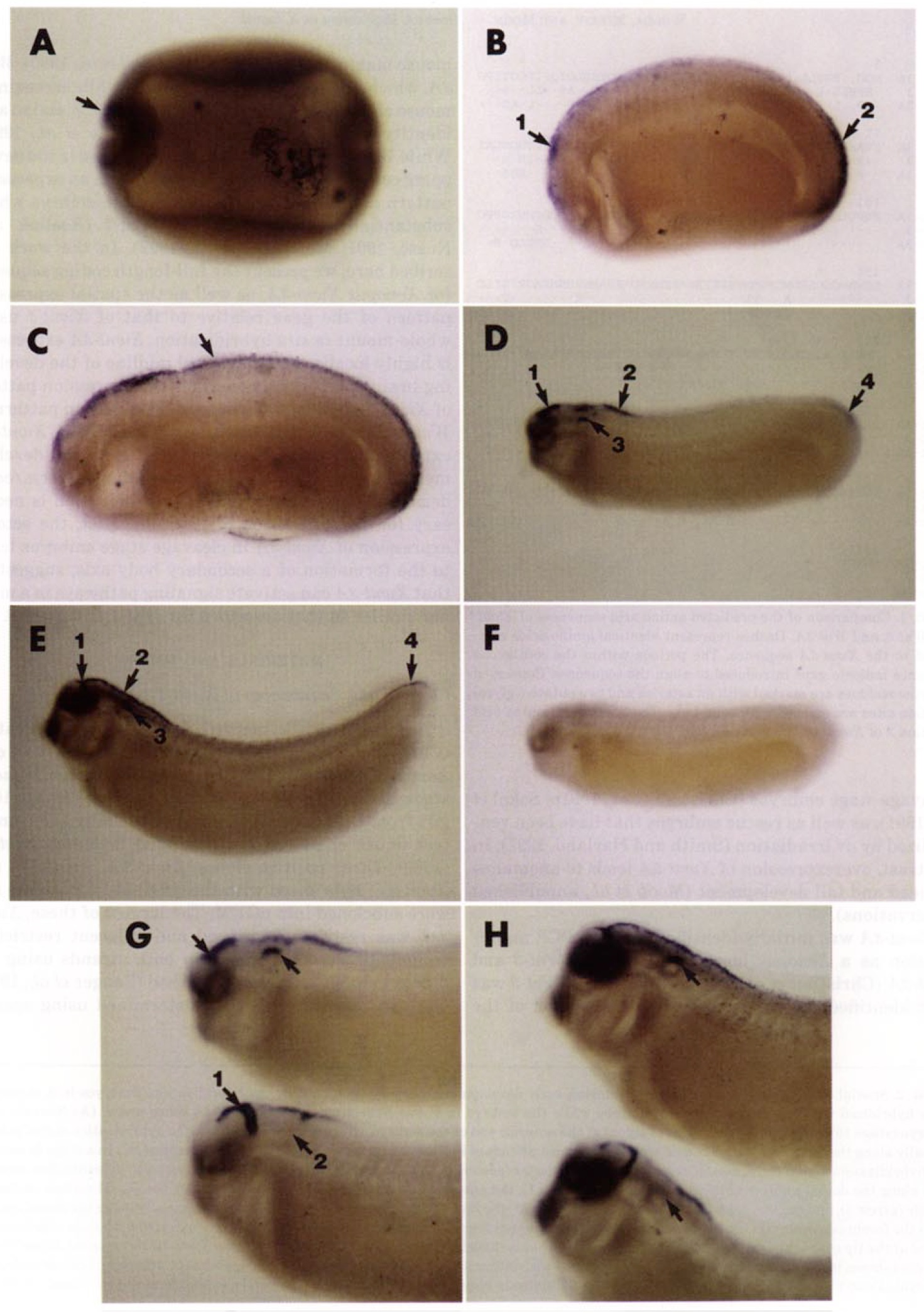
FIG. 2. Spatial expression pattern of Xwnt-3A during early development using whole-mount in situ hybridization. Embryos in A through E were hybridized with an Xwnt-3A antisense probe while the embryo in F was hybridized with an Xwnt-SA sense probe. (A) Neurula stage embryo (stage 16). Arrow indicates patch of signal at the anterior end of the embryo. (B) In a stage 22 embryo, the hybridization signal extends dorsally along the anterior/posterior axis and appears most abundant in the head (arrow 1) and tail (arrow 2) regions. (C) In a stage 24 embryo, the hybridization signal continues to extend along the anterior/posterior axis (arrow). (D) By stage 27 (hatching), the hybridization signal is seen along the dorsal midline of the mesencephalon (arrow 1), the anterior portion of the neural tube (arrow 2), the dorsal surface of the otic vesicle (arrow 3), and the tip of the tail (arrow 4). (E) At stage 31, theXwnt-3A hybidization signal extends continuously along the dorsal midline from the forebrain/ midbrain junction (arrow 1) to the anterior portion of the neural tube (arrow 2) and is still detected in the otic vesicle (arrow 3) and at the tip of the tail (arrow 4). (F) A stage 27 embryo has no hybridization signal when probed with an Xwnt-3A sense probe. Stage 27 or 31 embryos shown in G and H, respectively, were hybridized with either an Xwnt-3A antisense probe (top embryo) or an Xwnt-1 antisense (bottom embryo). Xwnt-1 is uniquely expressed at the midbrain/ hindbrain boundary (arrow 1) and is completely absent from the otic vesicle (arrow 2).
Image published in: Wolda SL et al. (1993)
Copyright © 1993. Image reproduced with permission of the Publisher, Elsevier B. V.
| Gene | Synonyms | Species | Stage(s) | Tissue |
|---|---|---|---|---|
| wnt3a.S | wnt-3a, Xwnt-3a, xwnt3a | X. laevis | Sometime during NF stage 16 to NF stage 31 | neural tube hindbrain |
| wnt1.L | int-1, int1, wnt-1, Xint-1, Xwnt-1, Xwnt1 | X. laevis | Throughout NF stage 27 to NF stage 31 | hindbrain |
Image source: Published
Permanent Image Page
Printer Friendly View
XB-IMG-138946