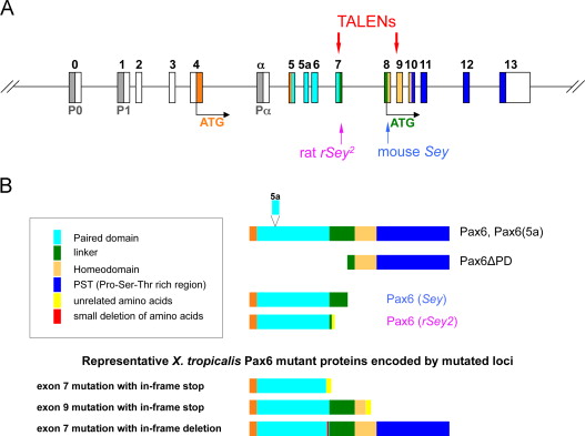
Fig. 1. Strategy for targeted mutagenesis of Xenopus tropicalis pax6 locus. (A) Structure of pax6 gene is shown schematically (not to scale). P0, P1, Pα are promoters (shaded in grey). Exons (boxes) are numbered or named on top (0 to 13 or α) and color coded: white, untranslated regions (UTRs); orange, N-terminal region before the paired domain; other colors correspond to color codes for protein domains shown in (B). Two translation start sites are shown (ATG): orange, for full-length isoforms (Pax6 with or without 5a); green, for paired-less isoform (Pax6δPD). Positions of mouse Sey and rat rSey2 mutations are indicated by arrows on the bottom. Two pairs of TALEN targets in exon 7 and exon 9 are shown on top. (B) Structures of Pax6 wild-type and representative mutant proteins are shown schematically (not to scale). Color codes of protein domains are shown in the box except the first three amino acids, which are shown in orange. Note that the paired domain is located between amino acids 4â131 and the homeodomain between amino acids 212â272 in X. tropicalis due to insertion of two amino acids between the two domains. The amino acid sequence of each domain is as shown in Walther and Gruss (1991). Most but not all mutations have some non-Pax6 amino acids at the C-terminus. Note that X. tropicalis mutations in exon 7 could theoretically encode Pax6δPD if mutations were not simultaneously introduced in exon 9 (not shown in the drawing). (For interpretation of the references to color in this figure legend, the reader is referred to the web version of this article.)
Image published in: Nakayama T et al. (2015)
Copyright © 2015. Image reproduced with permission of the Publisher, Elsevier B. V.
Permanent Image Page
Printer Friendly View
XB-IMG-140009