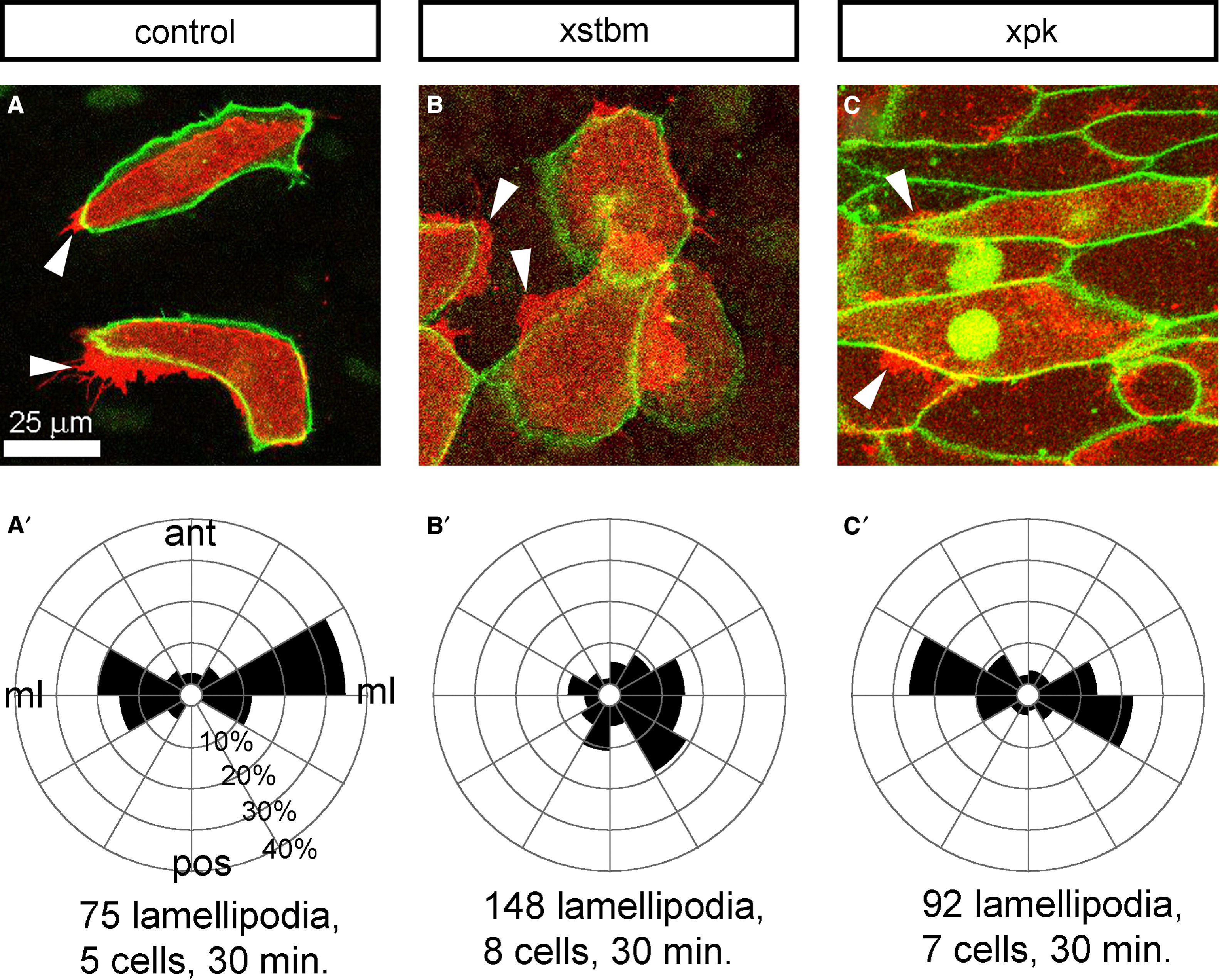
Figure 3. Cell Protrusions along the Fibronectin Substrate in Explants Overexpressing Xpk or Xstbm Xpk or Xstbm mRNA was injected into two dorsal blastomeres of the 4-cell-stage embryo. An mRNA encoding a plasma membrane localizing GFP (gap43-GFP) was injected into several dorsal blastomeres at stage 7 to allow visualization of cell protrusions and cell shapes. Dorsal open-faced explants were dissected from the injected embryos at stage 10 and cultured on the fibronectin-coated glass exposing the deep cells to confocal time-lapse imaging. The protrusions were detected on the surface on labeled cells facing the fibronectin substrate (red) and cell shapes were observed 5 μm deeper into the explant (green). (A) In control explants, gap43-GFP-labeled cells adopt bipolar shapes with protrusions at their mediolateral ends. (B) Cells in Xstbm-overexpressing explants remain isodiametric, do not adopt bipolar shapes, and extend protrusions in all directions. (C) Cells in Xpk-overexpressing explants show similar bipolar shapes and mediolaterally directed protrusions. Arrowheads show lamelliform protrusions along the fibronectin substrate. (A′, B′, and C′) Analysis of protrusive activity in (A), (B), and (C), respectively. Rose diagrams show the normalized frequency of protrusions from five cells in the explant shown in (A), eight cells in the explant shown in (B), and seven cells shown in the explant shown in (C) directed into 30° bins representing 360° around the cell’s center.
Image published in: Goto T et al. (2005)
Copyright © 2005. Image reproduced with permission of the Publisher, Elsevier B. V.
Permanent Image Page
Printer Friendly View
XB-IMG-144949