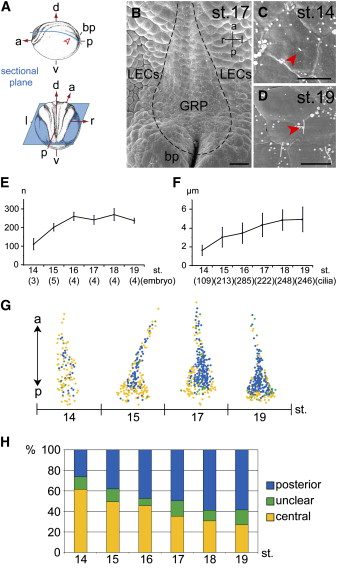
Figure 1. Characterization of Monocilia on the Gastrocoel Roof Plate of Xenopus Embryos(A) Schematic representation of dissection of a neurula frog embryo at stage 14. Abbreviations are as follows: a, anterior; bp, blastopore; d, dorsal; l, left; p, posterior; r, right; and v, ventral.(B) Scanning electron micrograph of stage (st.) 17 explant in which the gastrocoel roof plate (GRP) is indicated by a dashed line. Please note the distinct appearance of GRP cells that are organized in a flat epithelium, are monociliated, and possess a surface area 2.5 times smaller than that of the neighboring lateral endodermal cells (LECs). The scale bar represents 50 μm.(C and D) Central localization of a short cilium at stage 14 (C) and posterior localization of a long cilium at stage 17 (red arrowheads). The scale bar represents 10 μm.(E) The number (±SD) of ciliated GRP cells increases from about 100 at stage 14 to a plateau of 240â270 at stage 16â19.(F) The length of cilia (±SD) increases from 1.6 μm at stage 14 to 5 μm at stage 18/19.(G) Unequal distribution of central (yellow) and posteriorly polarized (blue) cilia across the GRP from stage 14â19. Cilia that could not be evaluated are indicated in green. Note that ordered distribution of polarized cilia starts at stage 15 and that the posterior and lateral parts of the GRP display mostly central cilia (cf. Figure S2).(H) Increase in the contingent of polarized cilia over time.
Image published in: Schweickert A et al. (2007)
Copyright © 2007. Image reproduced with permission of the Publisher, Elsevier B. V.
Permanent Image Page
Printer Friendly View
XB-IMG-153550