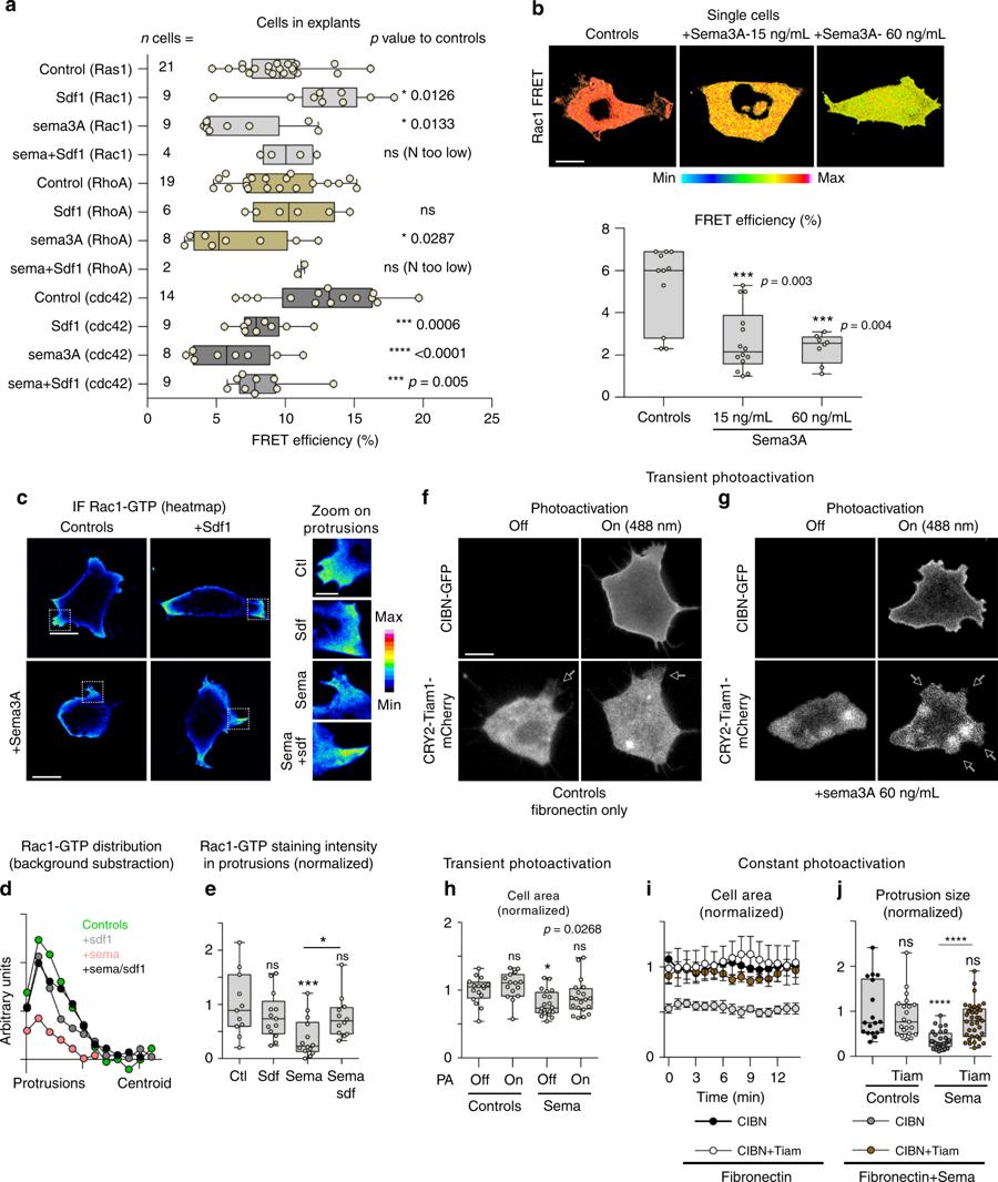
fig. 6. Rac1 activation is sufficient to rescue exposure to Sema3A. a Rac1, RhoA and Cdc42 activity assessed by FRET in cells from explants cultured in control, Sdf1 and/or Sema3A conditions, n = 118 cells (Rac1: controls(21), Sdf1(9), Sema3A(9), Sema3A + Sdf1(4); RhoA: controls(19), Sdf1(6), Sema3A(8), Sema3A + Sdf1(2); Cdc42: controls(14), Sdf1(9), Sema3A(8), Sema3A + Sdf1(9)) from three independent experiments. For each FRET probe, Sdf1, Sema3A and Sdf1+Sema3A conditions were compared to their cognate controls via ANOVA followed by multiple comparisons, individual p values are indicated on the figure. b Rac1 FRET in single cells under control conditions (Fibronectin) or with Fibronectin plus Sema3A coated at 15 or 60 ng/mL, n = 33 cells (ncontrols = 11, nSema3A-15ng.mL = 14 and nSema3A-60ng.mL = 8), ANOVA followed by multiple comparisons, p values indicated on the graph. c Immunofluorescence against Rac1-GTP. d Distribution of rac1 intensity from cell protrusions to cell centroid. Rac1 staining intensity in the cell centroid was measured in each condition and subtracted from each data set, n = 50 cells from one experiment. e Rac1 staining intensity in protrusions for each experimental condition, n = 50 protrusions from one experiment, ANOVA, followed by multiple comparisons; *p < 0.05; ***p < 0.001. f Photoactivation experiment with single cells transfected with CIBN-Caax-GFP and Tiam1-CRY2-mCherry under control conditions. g Photoactivation experiments with single cells transfected with CIBN-Caax-GFP and Tiam1-CRY2-mCherry cultured on Fibronectin, plus Sema3A coated at 60 ng/mL. h Normalised cell area for experimental conditions displayed in f and g, n = 71 cells (ncontrols-PA/OFF = 16, ncontrols-PA/ON = 15, nSema3A-PA/OFF = 20, and nSema3A-PA/ON = 20) from seven independent experiments, ANOVA followed by multiple comparisons, p value indicated on the graph. i Cell area overtime (mean+s.e.m) for cells under sustained photoillumination after being transfected with CIBN and Tiam on Fibronectin or Fibronectin plus Sema3A coated at 60 ng/mL or cells transfected with CIBN only on Fibronectin plus Sema3A coated at 60 ng/mL, n = 21 cells from one experiment (nCIBN/FN = 3, nCIBN+Tiam/FN = 4, nCIBN/Sema = 8, and nCIBN+Tiam/Sema = 6). j Size of protrusions from cells used in f and g, n = 105 protrusions (nCIBN/FN = 18, nCIBN+Tiam/FN = 21, nCIBN/Sema = 29, nCIBN+Tiam/Sema = 37). ANOVA followed by multiple comparisons, ****p < 0.0001. Scale bars 10 μ, except for zooms in panel c, 3 μ. Box and whiskers plot: the box extends from the 25th to the 75th percentile; the whiskers show the extent of the whole dataset. The median is plotted as a line inside the box. Source data are provided as a Source Data file
Image published in: Bajanca F et al. (2019)
© The Author(s) 2019. Creative Commons Attribution license
Permanent Image Page
Printer Friendly View
XB-IMG-175704