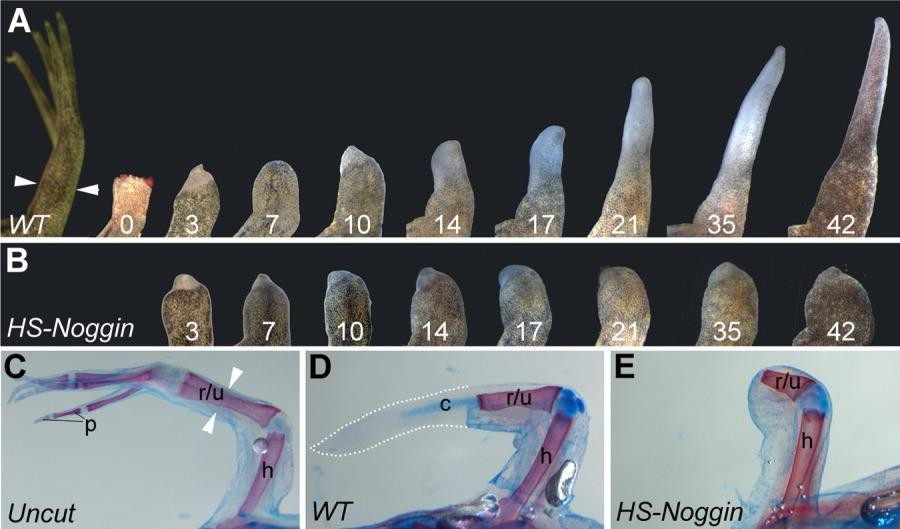
Figure 4. Regeneration of the froglet forelimb. A: A normal froglet forelimb has 4 digits (far left). Amputation of the Xenopus froglet forelimb midway along the forearm (zeugopod, white arrowheads) results in epimorphic regeneration of a hypomorphic spike. Regenerative progress is shown at 0 (time of cutting) to 42 days afterward. From days 0-3, soft tissue contracts away from the wound surface and the wound becomes covered. A blastema is present by day 7 and extends distally forming a spike. B: In transgenic animals in which the Bmp signalling pathway is inhibited by induction of ectopic noggin expression with daily heat shocks over 14 days, regeneration of the spike is completely inhibited. C: Skeletal preparation of a normal froglet forelimb showing the ossified bone in red and the cartilage in blue (alizarin red S/alcian blue). The white arrowheads show the position of amputation. D: Skeletal preparation of normal wild-type regenerating forelimb after 42 days. The extent of the regenerated spike is outlined by white dots. Only cartilage is present in the spike, and there are no joints. E: Bmp inhibited animals show no regeneration of cartilage or other soft tissues. c, cartilage; h, humerus; r/u, radius and ulna; p, phalanges.
Image published in: Beck CW et al. (2009)
Copyright © 2009. Image reproduced with permission of the Publisher, John Wiley & Sons.
Permanent Image Page
Printer Friendly View
XB-IMG-26348