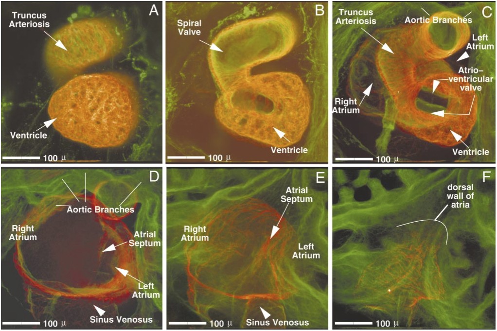
FIG. 5. Ventral-to-dorsal progression through a stage 46 Xenopus heart. Images were digitally created from a series of 48 optical sections (taken 5um apart) through a double-labeled stage 46 Xenopus heart (red, CT3; green, JB3). Image A represents the most ventral 8 images of this series, digitally merged; image B, the next 8, etc. The relative positioning of heart structures to one another, as well as an individual structure order of emergence within the series, can be established: the truncus arteriosus and ventricle are always most ventral (A). Progressing dorsally, the spiral valve (B) appears within the truncus. Next, the single atrioventricular valve (C) is seen overlying the atrial septum (D and E). Midway through the series (C and D), the first views of the atria, as well as the aortic branches, are viewed. Both atria and the sinus venosus are seen more dorsally (D and E). Finally, the back wall of the atria is seen, positioned most dorsal (F). Scale bar, 100 um.
Image published in: Kolker SJ et al. (2000)
Copyright © 2000. Image reproduced with permission of the Publisher, Elsevier B. V.
| Gene | Synonyms | Species | Stage(s) | Tissue |
|---|---|---|---|---|
| tnnt2.L | CT3, LOC446916 | X. laevis | Throughout NF stage 46 | heart muscle myocardium left atrium right atrium cardiac ventricle spiral septum aorta pulmonary artery |
| fbn2.L | JB3 | X. laevis | Throughout NF stage 46 | heart left atrium right atrium atrioventricular valve leaflet endocardium spiral septum aortic arch aorta pulmonary artery |
Image source: Published
Permanent Image Page
Printer Friendly View
XB-IMG-48757