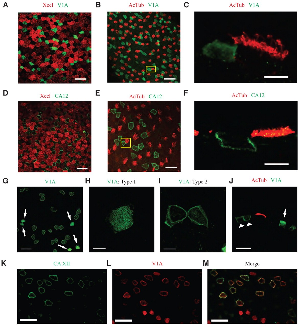
Fig. 2. Most ionocytes develop in contact with ciliated cells. (AâF) Whole-mount co-immunostaining for xeel (A,D), acetylated α-tubulin (B,E) and v1a (AâC) or ca12 (DâF) in X. tropicalis tadpole epidermis shows that v1a-or ca12-expressing cells are neither goblet or ciliated cells but develop in close association with ciliated cells. High magnification of the area enclosed in a yellow box in B and E is shown in cross-section in C and F. v1a (C; green) shows close coupling with ciliated cells (C; red), whereas ca12 shows distinct basolateral staining in these transverse sections (F; green). The ca12-expressing cell (green) is also in direct contact with a ciliated cell (red). (G) Immunostaining of v1a (green) shows two distinct cell types. Approximately 30% of cells show bright apical staining corresponding to type 1 cells (highlighted with arrows), whereas the remaining v1a-expressing cells (type 2) show lateral membrane staining. (H,I) High magnification of type 1 and type 2 cells, respectively. (J) Cross-section of type 1 (arrow) and type 2 (arrowhead) v1a-expressing cells (green) shown adjacent to ciliated cells (red). (KâM) Co-immunostaining of ca12 (green) with v1a (red) shows colocalisation of these two markers in type 2 cells. Scale bars: 50 μm (A,B,D,E,G,KâM); 10 μm (C,F,H,I); 20 μm (J).
Image published in: Dubaissi E and Papalopulu N (2011)
© 2011. Creative Commons Attribution-NonCommercial-ShareAlike license
| Gene | Synonyms | Species | Stage(s) | Tissue |
|---|---|---|---|---|
| itln1 | hl-1, intl, itln, lfr, omentin, xeel | X. tropicalis | Throughout NF stage 33 and 34 | epidermis non-cililated epidermal cell ciliated epidermal cell ciliated cell |
| tuba4a.L | alpha tubulin, alpha-tubulin, tuba4, tuba4b | X. laevis | Throughout NF stage 33 and 34 | epidermis ciliated epidermal cell ciliated cell |
| atp6v1a.L | atp6a1, atp6v1a1, atpv1a, ho68, v1a, va68, vma1, vpp2 | X. laevis | Throughout NF stage 33 and 34 | epidermis non-cililated epidermal cell |
| ca12.L | X. laevis | Throughout NF stage 33 and 34 | epidermis non-cililated epidermal cell |
Image source: Published
Permanent Image Page
Printer Friendly View
XB-IMG-74401