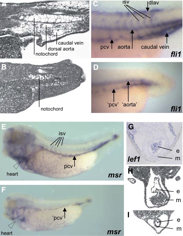
Fig. 5. The cardiovascular system is affected by lef1 depletion. Sagittal section of a stage 38 control embryo (A) showing the dorsal aorta and the caudal vein. In lef1 depleted embryos (B), the tail lacks the major blood vessels, the caudal vein and the dorsal aorta. Fli1 expression in the endothelium of a control embryo (C). isv, intersomitic vessels. dlav, dorsal longitudinal anastomosing vessel. pcv, posterior cardinal vein. aorta, dorsal aorta. Lef1 depleted embryos show strongly reduced expression of fli1 (D) at the position of the pcv and the aorta and no indication of intersomitic vessels, dlav or caudal vein. Msr/APJ expression in control embryo (E) and in lef1 depleted embryo (F) showing reduced expression. Endogenous expres- sion of lef1 in the heart of a stage 34 control embryo (G). e, endocardium. m, myocardium. Transverse section of the heart of a control embryo (H) and of a lef1 depleted embryo (I) which shows severe retardation of heart development both for the endocardium and myocardium.
Image published in: Roel G et al. (2009)
Copyright © 2009. Image reproduced with permission of the Publisher, University of the Basque Country Press.
| Gene | Synonyms | Species | Stage(s) | Tissue |
|---|---|---|---|---|
| lef1 | lef-1, xlef-1, xlef1 | X. tropicalis | Throughout NF stage 33 and 34 | myocardium endocardium heart |
| fli1 | fli, fli-1, fli1-a, Xfli, Xfli-1, Xfli1 | X. tropicalis | Throughout NF stage 37 and 38 | blood vessel dorsal longitudinal anastomosing vessel posterior cardinal vein caudal vein aorta intersomitic vessel |
| aplnr | agtrl1, angio1, apj, apjr, aplnr-a, aplnr-b, hg11, msr, Xangio1, X-msr, Xmsr | X. tropicalis | Throughout NF stage 37 and 38 | posterior cardinal vein intersomitic vessel heart tail bud infundibulum |
Image source: Published
Permanent Image Page
Printer Friendly View
XB-IMG-75482