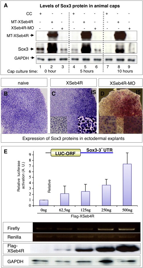
Fig. 5. XSeb4R strongly activates maternal Sox3 translation. (A) Western blot analysis of Sox3 translation in caps from MT-XSeb4R or XSeb4R-MO injected embryos, using an anti-Sox3 antibody. GAPDH antibody was used as loading control. Note a progressively increased amount of Sox3 signal in MT-XSeb4R overexpressing caps at the equivalent of blastula, t0 (lane 2) and gastrula, t5 (lane 5), but not at neurula, t10 (lane 8) stages of explant development. Observe a decrease in the level of Sox3 signal at the time of explant dissection (t0) in XSeb4R depleted caps (lane 3) compared to the control (lane 1). (BâD) Ectodermal explants dissected at the blastula or gastrula stage from embryos that were injected unilaterally with XSeb4R mRNA or XSeb4R-MO were stained with an anti-Sox3 antibody. Caps from control uninjected embryos show a uniform staining pattern (B; 100%, n = 50); expanded signals are observed on the injected side of explants from XSeb4R-overexpressing embryos (C; 100%, n = 50). Sox3 protein translation was not significantly affected in XSeb4R-depleted caps (D; 100%, n = 50; high magnification: compared signals in the cells stained in red and unstained areas). (E) Sox3-3â²UTR Luciferase reporter was analyzed in HEK293 cells. All firefly luciferase values were normalized to renilla luciferase. Shown are the relative luciferase expression activation; note that XSeb4R stimulates LUC-Sox3-3â²UTR in a concentration dependent manner. Four independent transfections revealed similar trends. Also observe increased firefly but not renilla luciferase mRNA stability in response to XSeb4R, as tested by RT-PCR. The increased amount of Flag-XSeb4R protein in transfected cells is added, with GAPDH as a protein loading control.
Image published in: Bentaya S et al. (2012)
Copyright © 2012. Image reproduced with permission of the Publisher, Elsevier B. V.
Permanent Image Page
Printer Friendly View
XB-IMG-77512