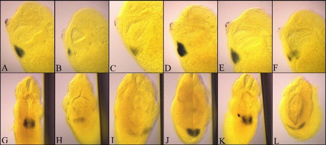
Fig. 4. Blocking RA signaling is required in a narrow window of time for formation of the heart tube. The heart has been visualized by whole-mount in situ hybridization for cTnI and is viewed in whole cleared embryos in side view (top panel) and from the anterior end (bottom panel) in order to visualize the formation of a tube. Normal heart tube formation can be seen in stage 31/32 embryos that were treated with 2 μl/ml DMSO as a carrier control (A, G). Treatment with 1 μM RA at stage 14 resulted in a dramatic decrease of myocardial differentiation (B, H). Treatment with 1 μM AGN 194301 resulted in a block to heart tube formation remained as a sheet along the ventral midline (C, I). cTnI expression levels and spatial distribution were restored if RA and AGN 194301 were given in equimolar amounts (D, J). When embryos were subjected to an initial treatment of 1 μM AGN 194301 at stage 14 with subsequent addition of 1 μM RA at stage 16, cTnI expression (E, K) was also similar to control patterns. However, when the addition of RA was delayed until stage 18/20, cardiomyocyte differentiation was shown to be again restricted to posterior regions of the normal heart field and linear heart tube formation was not detected (F, L). Thus, blocking RA signaling in a tight window of time between stages 14 and 18 is sufficient to prevent formation of the heart tube.
Image published in: Collop AH et al. (2006)
Copyright © 2006. Image reproduced with permission of the Publisher, Elsevier B. V.
Permanent Image Page
Printer Friendly View
XB-IMG-83515