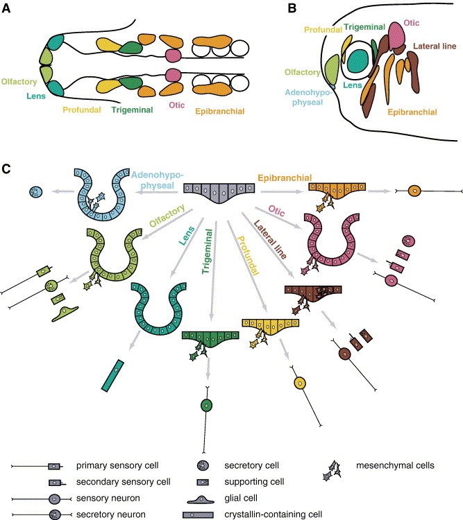
Fig. 1. Cranial placodes in vertebrates. (A) Cranial placodes in a 10- to 13-somite-stage chick embryo (modified from Streit, 2004; based on D'Amico-Martel and Noden, 1983; Bhattacharyya et al., 2004). In amniotes, profundal and trigeminal placode are commonly referred to as ophthalmic and maxillomandibular placode of the trigeminal nerve, respectively. (B) Cranial placodes in a tailbud stage Xenopus embryo (modified from Schlosser and Northcutt, 2000). (C) Schematic summary of morphogenesis and cellular derivatives of various cranial placodes. Invagination occurs in adenohypophyseal, olfactory, lens, and otic placodes. Moreover, in all placodes except the lens placode, some cells migrate away from the placodal epithelium as mesenchymal cells to form sensory neurons, secretory cells, or glial cells. In lateral line placodes, another subset of cells migrates along the basement membrane and forms the lateral line primordia (modified from Schlosser, 2005).
Image published in: Schlosser G (2006)
Copyright © 2006. Image reproduced with permission of the Publisher, Elsevier B. V.
Permanent Image Page
Printer Friendly View
XB-IMG-84068