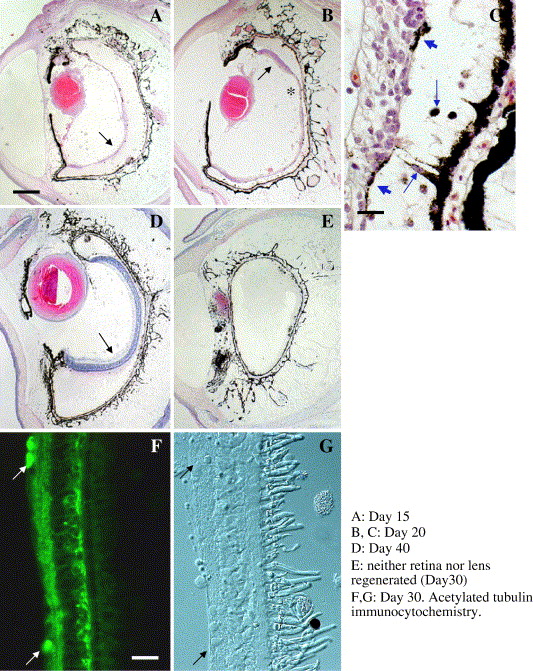
Fig. 4. Later stage of retinal regeneration in X. laevis eyes (4 to 5 months after metamorphosis). (A) Day 15, (B, C) Day 20, (D) Day 40 and (E) Day 30 after retinectomy. (A) By Day 15, lens structures are well recognized with intense eosin staining. Regenerating retinas (arrows) are still thin and are separated from RPE layer. (B) By Day 20, the laminar structure of the regenerating retina is partially developed at the periphery (arrow). The area indicated by asterisk is shown in (C) at a higher magnification. (C) A few RPE cells are found in the space between RPE layer and the retinal epithelium as shown by blue arrows. Some cells extend from RPE layer to the retinal layer (indicated by lower blue arrow). Blue-colored arrowheads indicate pigmented cells attached to the retinal layer. (D) By Day 40 a well-stratified retinal structure develops as shown by an arrow. (E) In some cases neither the retina nor lens regenerates, the eye cavity being occluded by connective tissue derived from the choroid tissue. (F, G) Laminar structure of the regenerating retina at Day 30 is well identified with acetylated tubulin staining. Arrows indicate immunoreactive ganglion cell bodies. G is a Nomarsky differential image of the same area. Scale bar in A is 200 μm and is applied to B, D, E. Scale bars in C and F are 20 μm.
Image published in: Yoshii C et al. (2007)
Copyright © 2007. Image reproduced with permission of the Publisher, Elsevier B. V.
| Gene | Synonyms | Species | Stage(s) | Tissue |
|---|---|---|---|---|
| tuba4a.L | alpha tubulin, alpha-tubulin, tuba4, tuba4b | X. laevis | Sometime during NF stage 65 to NF stage 66 | retina retinal ganglion cell |
Image source: Published
Permanent Image Page
Printer Friendly View
XB-IMG-149230