Click here to close Hello! We notice that you are using Internet Explorer, which is not supported by Xenbase and may cause the site to display incorrectly. We suggest using a current version of Chrome, FireFox, or Safari.
XB-IMG-24842

Xenbase Image ID: 24842
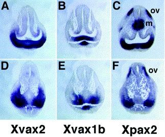

Fig. 2. Expression of Xvax2 (A,D), Xvax1b (B,E) and Xpax2 (C,F) as detected in transverse sections of stage 23 Xenopus embryos following whole-mount in situ hybridization, cut at the level of the anterior (A–C) or posterior (D–F) optic vesicle. m, mesencephalon; ov, otic vesicle.

Image published in: Liu Y et al. (2001)

Copyright © 2001. Image reproduced with permission of the Publisher, Elsevier B. V.

GeneSynonymsSpeciesStage(s)Tissue
vax2.Lxvax2, vax3, dres93, vax2-a, vax2-bX. laevisThroughout NF stage 23ventral
forebrain
optic vesicle
pax2.LXPax2, XPax-2, pax-2, pax2-a, pax2-b, LOC108697493X. laevisThroughout NF stage 23otic vesicle
vax1.Lvax, vax1b, vax1b-a, xvax1b, xvax1, vax1-b, vax1-aX. laevisThroughout NF stage 23optic vesicle
ventral
forebrain

Image source: Published

Larger Image
Printer Friendly View

Quality:
Good quality Poor quality
comment [+]
Return to previous page