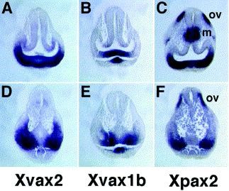
Fig. 2. Expression of Xvax2 (A,D), Xvax1b (B,E) and Xpax2 (C,F) as detected in transverse sections of stage 23 Xenopus embryos following whole-mount in situ hybridization, cut at the level of the anterior (AâC) or posterior (DâF) optic vesicle. m, mesencephalon; ov, otic vesicle.
Image published in: Liu Y et al. (2001)
Copyright © 2001. Image reproduced with permission of the Publisher, Elsevier B. V.
| Gene | Synonyms | Species | Stage(s) | Tissue |
|---|---|---|---|---|
| vax2.L | dres93, vax2-a, vax2-b, vax3, xvax2 | X. laevis | Throughout NF stage 23 | ventral forebrain optic vesicle |
| vax1.L | vax, vax1-a, vax1-b, vax1b, vax1b-a, xvax1, xvax1b | X. laevis | Throughout NF stage 23 | optic vesicle ventral forebrain |
| pax2.L | LOC108697493, pax-2, pax2-a, pax2-b, XPax-2, XPax2 | X. laevis | Throughout NF stage 23 | otic vesicle |
Image source: Published
Permanent Image Page
Printer Friendly View
XB-IMG-24842