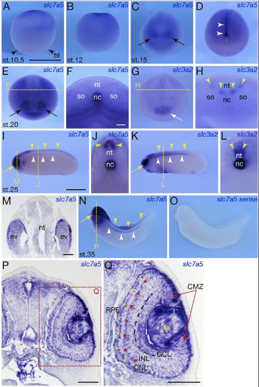
Fig. 3.Spatial expression of slc7a5.S and slc3a2 in the developmental stages. Whole-mount in situ hybridization was performed to analyze spatial expression ofslc7a5.S(shown in AâF, I, J, MâQ) and slc3a2(shown inG, H, K and L) in the developmental stages. (A)st.10.5. Lateral view. slc7a5.S expressed in theanimal hemisphere. Black arrowheads indicatethe blastopore lip (bl). (B) st.12. Lateral view.slc7a5.S expression was observed throughoutthe animal hemisphere at st.10.5. (C, D) st.15.(C) Anterior view.slc7a5.Slocalized at the eyeanlagen (black arrows). (D) Posterior view.slc7a5.Swas detected in the notochord from theblastopore to anterior along midline as indicatedby white arrowheads. (EâH) st.20. (E) Anteriorview. More definite expression ofslc7a5.Swasdetected at the eyefield in comparison to st.15embryo in (C) (black arrows). A yellow brokenline indicates the plane of section in panel (F).(F) Transverse section of st.20 embryo shown inpanel (E). Strong expression ofslc7a5.Swasobserved in the notochord (nc). (G) Anteriorview. Weak expression ofslc3a2was detected inthe cement grand (white arrow). A yellowbroken line indicates the plane of section inpanel (H). (H) Transverse section of st.20 em-bryo shown in panel (G). Spotted expression ofslc3a2was observed in the neural tube (yellowarrowheads), but no expression was detected inthe notochord unlikeslc7a5.Sexpression in st.20embryo. (IâL) st.25. (I) Lateral view.slc7a5.Sexpression was intensely detected in the eye(yellow arrow) and also observed in the neuraltube (yellow arrowheads) and the notochord(white arrowheads). Two yellow broken linesrepresent the plane of section in panel (J) and(M). (J) Transverse section of st.25 embryo attrunk region shown in panel (I).slc7a5.Swasobserved in the dorsal part of the neural tube(nt, yellow arrowheads) and the notochord (nc).(K) Lateral view.slc3a2expression was intenselydetected in the eye (yellow arrow) and also ob-served in the neural tube (yellow arrowheads)and the notochord (white arrowheads) asslc7a5.Sexpression shown in panel (I). (L)Transverse section of st.25 embryo shown inpanel (K).slc3a2was observed in the ventralpart of the neural tube (nt, yellow arrowheads)and the notochord (nc). (M) Transverse sectionof st.25 embryo at trunk region shown in panel(I).slc7a5.Sexpressed in the eye vesicle (ev) andthe neural tube (nt). (NâQ) st.35. (N) Lateralview.slc7a5.Sexpression was maintained in theeye (yellow arrow), the neural tube (yellow ar-rowhead) and the notochord (white arrowhead).A yellow broken line indicates the plane of sec-tion in panel (P). (O) Negative control with a sense probe ofslc7a5.S. (P) Transverse section of st.35 embryo. A rectangle with red broken line represents a regionmagnified in panel (Q). (Q) Higher magnification of photomicrograph of rectangle (Q) in panel (P).slc7a5.Sexpressed in the lens (le), CMZ of the neural retina (redarrow),the border of INL and ONL (red asterisks) and RPE (yellow asterisks). Scale bars: 1 mm in AâE and G; 0.1 mm in F, H, J and L; 1 mm in I, K, N and O; 0.1 mm inM; 0.1 mm in P; 0.1 mm in Q. bl: blastopore lip, CMZ: ciliary marginal zone of the neural retina, ev: eye vesicle, nc: notochord, nt: neural tube, so: somite, GCL:ganglion cell layer, INL: inner nuclear layer, le: lens, ONL: outer nuclear layer, RPE: retinal pigment epithelium. (For interpretation of the references to color in thisfigure legend, the reader is referred to the web version of this article.)
Image published in: Katada T and Sakurai H (2019)
Copyright © 2019. Image reproduced with permission of the Publisher, Elsevier B. V.
| Gene | Synonyms | Species | Stage(s) | Tissue |
|---|---|---|---|---|
| slc7a5.S | 4f2lc, cd98, e16, L amino acid transporter-1, LAT-1, lat1, mpe16, slc7a5-a, slc7a5-b | X. laevis | Sometime during NF stage 10.5 to NF stage 12 | animal hemisphere |
| slc7a5.S | 4f2lc, cd98, e16, L amino acid transporter-1, LAT-1, lat1, mpe16, slc7a5-a, slc7a5-b | X. laevis | Throughout NF stage 15 | eye primordium notochord |
| slc7a5.S | 4f2lc, cd98, e16, L amino acid transporter-1, LAT-1, lat1, mpe16, slc7a5-a, slc7a5-b | X. laevis | Throughout NF stage 20 | optic vesicle notochord |
| slc3a2.L | 4f2, 4t2hc, nacae | X. laevis | Throughout NF stage 20 | cement gland primordium neural tube |
| slc7a5.S | 4f2lc, cd98, e16, L amino acid transporter-1, LAT-1, lat1, mpe16, slc7a5-a, slc7a5-b | X. laevis | Throughout NF stage 25 | optic vesicle notochord neural tube |
| slc3a2.L | 4f2, 4t2hc, nacae | X. laevis | Throughout NF stage 25 | optic vesicle neural tube notochord |
| slc7a5.S | 4f2lc, cd98, e16, L amino acid transporter-1, LAT-1, lat1, mpe16, slc7a5-a, slc7a5-b | X. laevis | Throughout NF stage 35 and 36 | eye spinal cord notochord lens ciliary marginal zone retinal inner nuclear layer retinal outer nuclear layer retinal pigmented epithelium brain |
Image source: Published
Permanent Image Page
Printer Friendly View
XB-IMG-174699