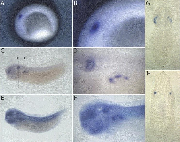
Fig. 2. Fox J1.2. Panels AâF show lateral views of embryos with the anterior to the left. Embryos were stained for FoxJ1.2 expression at stages 23 (A,B), 28 (C,D), and 35 (E,F). The second image is a closer view of the stained region. We sectioned and cleared stained stage 28 embryos to visualize staining in the otic vesicle (G) and pronephros (H). The sections correspond to the lines drawn in panel C and are shown with the dorsal side up.
Image published in: Choi VM et al. (2006)
Copyright © 2006. Image reproduced with permission of the Publisher, Elsevier B. V.
| Gene | Synonyms | Species | Stage(s) | Tissue |
|---|---|---|---|---|
| foxj1.2 | X. tropicalis | Throughout NF stage 23 | otic placode | |
| foxj1.2.L | X. laevis | Throughout NF stage 28 | otic placode pronephric mesenchyme | |
| foxj1.2.L | X. laevis | Throughout NF stage 35 and 36 | head otic vesicle pharyngeal arch |
Image source: Published
Permanent Image Page
Printer Friendly View
XB-IMG-3025