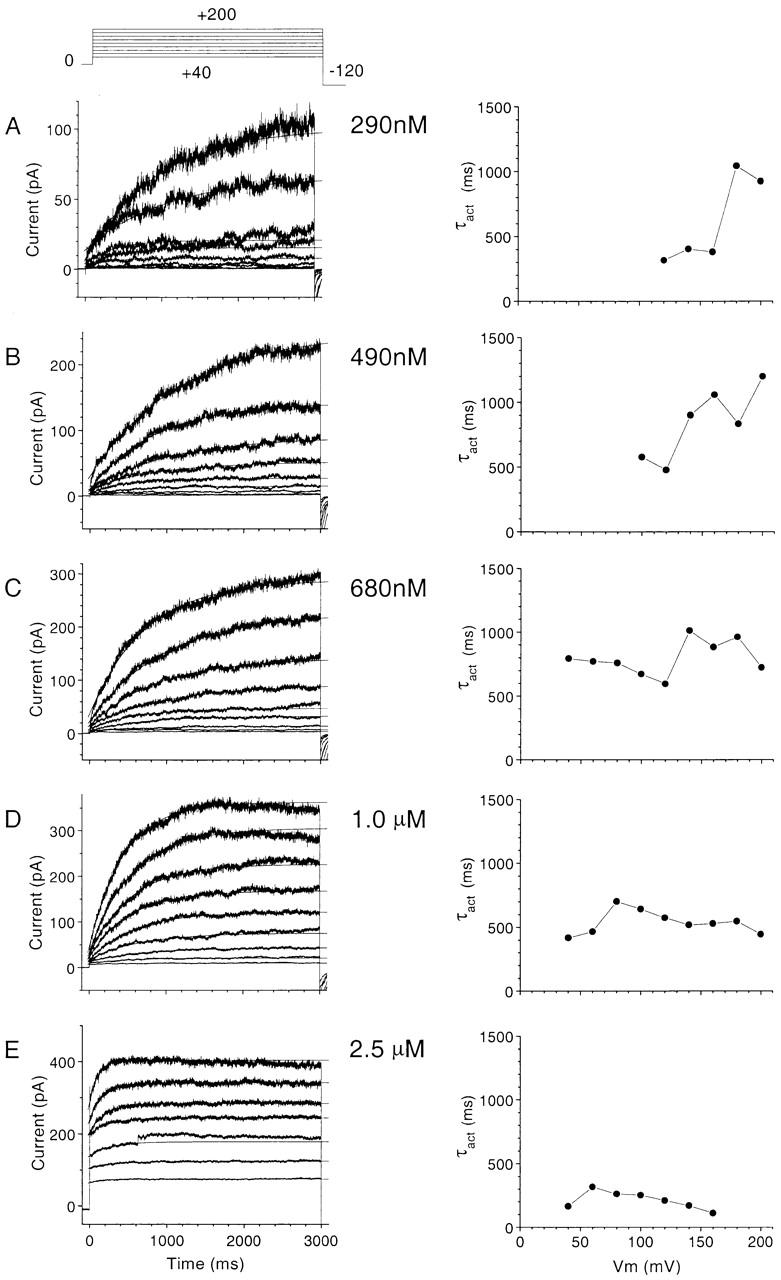
Figure 9. Kinetic analysis of activation of Ca2+-activated Cl− currents in a representative excised patch. Currents were elicited by voltage-clamp steps applied while the cytosolic face of the patch was exposed to solutions with different free [Ca2+]. The patch was voltage clamped by stepping to various potentials between +200 and +40 mV. The activating phase of the currents were fitted to single exponentials (superimposed) and the time constants were plotted versus membrane potential.
Image published in: Kuruma A and Hartzell HC (2000)
© 2000 The Rockefeller University Press. Creative Commons Attribution-NonCommercial-ShareAlike license
Permanent Image Page
Printer Friendly View
XB-IMG-117071