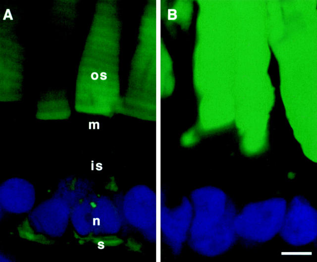
Figure 8. Overexpression of the fusion proteins is not the cause of delocalization to the RIS. Transgenic eyes expressing GFP-CT44 and GFP-CT44/P353L were excised, fixed, and sectioned in parallel. Frozen sections from each eye were consecutively imaged by confocal microscopy. All image acquisition settings were identical for both samples and postmicroscopy processing was done in parallel to the images. GFP-CT44 (A) was expressed at a much higher level than GFP-CT44/P353L (B) and yet was not found in significant levels in the RIS in contrast to the mutated fusion protein. GFP (green) and Hoescht 33342 (blue). os, outer segment; is, inner segment; n, nucleus; m, mitochondria; and s, synaptic terminal. Bar, 5 μm.
Image published in: Tam BM et al. (2000)
© 2000 The Rockefeller University Press. Creative Commons Attribution-NonCommercial-ShareAlike license
Permanent Image Page
Printer Friendly View
XB-IMG-118098