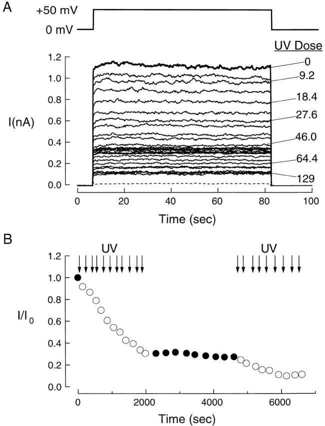
Figure 6. Effect of UV light on CNG channel currents. (A) Ionic currents before and after UV exposure. The currents were recorded in symmetrical 130-mM NaCl from an excised, inside-out membrane patch from a Xenopus oocyte membrane expressing bovine rod CNG (RET) channels. The template at top shows the timing of the voltage step. The bold trace at top is the current in the presence of 100 μM (approximately half-saturating) intracellular cGMP before UV exposure. The solid traces of successively smaller amplitude were obtained after increasing cumulative UV exposures. UV doses (in photons × 108 · μm−2) are indicated for selected traces. The patch was irradiated with light from a xenon arc lamp (280 nm) in the absence of cGMP. The light intensity was 4.6 × 10⁸ photons μm−2 s−1. The cyclic GMP-activated currents were corrected for leak by subtracting the current in the absence of cGMP after the same dose. The dashed trace shows the leak current before irradiation. The initial cGMP-activated current was 1,120 pA; the current at the end of the experiment (after a cumulative dose of 1.3 × 10¹⁰ photons · μm−2) was 125 pA. (B) Light and time dependence of currents. The channel current (I), normalized by the current before UV exposure (I0), is plotted as a function of time during the experiment. Each open symbol represents the current recorded after a 1-s exposure containing 4.6 × 10⁸ photons · μm−2 except for the last two open symbols, which were recorded after 5-s exposures that each contained 2.3 × 10⁹ photons · μm−2. Closed symbols denote currents recorded with no intervening UV exposure. Arrows indicate timing of UV applications. Results are from patch in A.
Image published in: Middendorf TR et al. (2000)
© 2000 The Rockefeller University Press. Creative Commons Attribution-NonCommercial-ShareAlike license
Permanent Image Page
Printer Friendly View
XB-IMG-121121