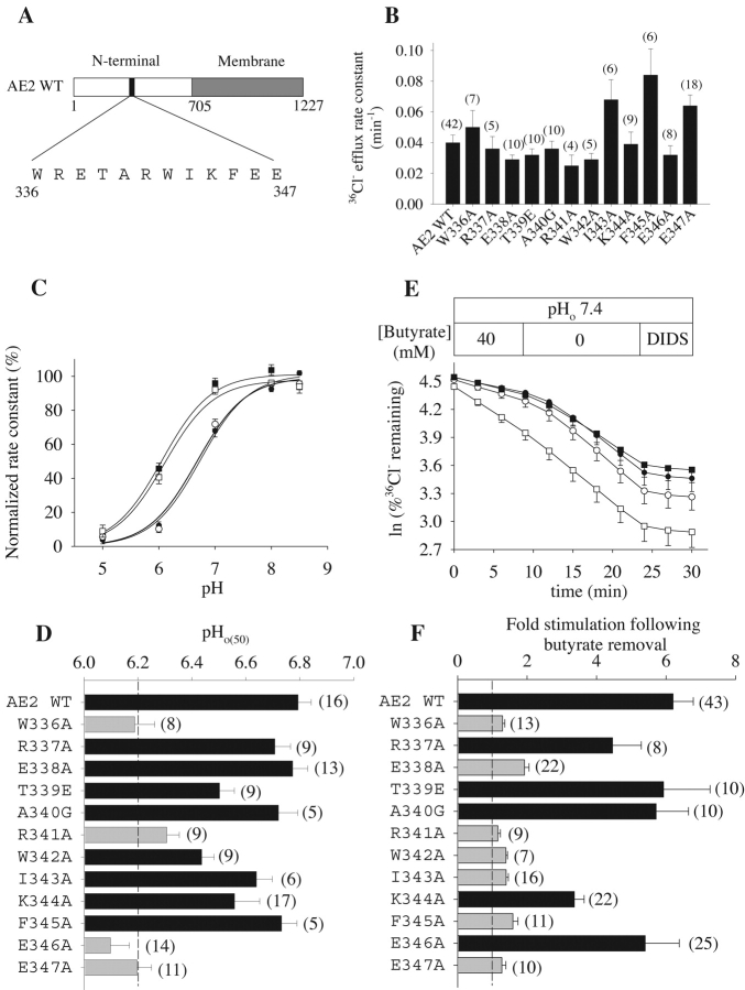
Figure 3. . Systematic point mutagenesis of AE2 residues 336–347 identifies amino acids important for regulation of Cl− transport by pHo and by pHi. (A) Schematic of point mutations in the NH2-terminal cytoplasmic domain of AE2. (B) 36Cl− efflux rate constants measured at pHo 7.4 in n oocytes expressing wild-type AE2 or the indicated AE2 substitution point mutants (mean ± SEM). (C) Regulation by pHo of normalized 36Cl− efflux from oocytes expressing wild-type AE2 (filled circles) or the AE2 mutants R337A (open circles), E346A (filled squares), and E347A (open squares). Values are means ± SEM. (D) pHo(50) values for the indicated single codon mutants (means ± SEM). Gray bars indicate pHo(50) values significantly different from wild-type AE2 (P < 0.05, Student's unpaired t test). (E) Representative time course of 36Cl− efflux from oocytes expressing wild-type AE2 (closed circles) or the AE2 mutants R337A (open circles), E346A (filled squares), or E347A (open squares) during elevation of pHi by removal of bath butyrate (40 mM) and subsequent inhibition by DIDS (200 μM). (F) Mean fold stimulation (±SEM) of Cl− efflux following bath butyrate removal from n oocytes expressing wild-type AE2 or the indicated AE2 point mutants. Gray bars indicate mutants for which stimulation values differed significantly from wild-type AE2 (P < 0.05, Student's unpaired t test).
Image published in: Stewart AK et al. (2002)
Copyright © 2002, The Rockefeller University Press. Creative Commons Attribution-NonCommercial-ShareAlike license
Permanent Image Page
Printer Friendly View
XB-IMG-121212