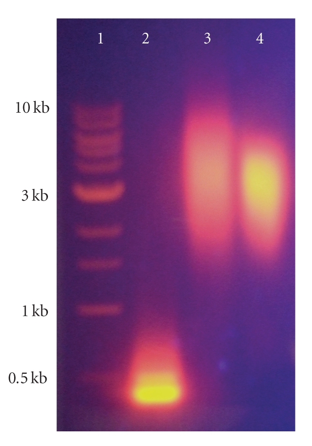
Figure 4. Agarose gel electrophoresis of qdots conjugates (1% agarose gel; Tris-acetate-EDTA buffer containing ethidium bromide for DNA visualization; 80 V potential difference). A 1-kb DNA ladder (Lane 1; New England Biolabs, Ipswich, Mass, USA), with DNA fragments ranging from 0.5–10 kilobases (kb) as indicated, was utilized to illustrate relative electrophoretic mobility of the qdot conjugates. Unconjugated AMP-coated qdots (Lane 2) have an increased mobility by comparison with both muscimol-conjugated qdots (Lane 3) and qdots conjugated with methoxy terminated PEG2000 (Lane 4), indicating successful functionalization of the qdot surface.
Image published in: Tomlinson ID et al. (2007)
Image downloaded from an Open Access article in PubMed Central. Copyright © 2007 Ian D. Tomlinson et al.
Permanent Image Page
Printer Friendly View
XB-IMG-122879