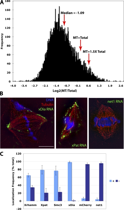
Figure 1. Specific mRNAs are enriched on X. laevis meiotic microtubules. mRNA from total X. laevis egg extract or from purified taxol-stabilized microtubules was hybridized to Affymetrix microarrays. The ratio of the signal of each microtubule-associated mRNA to its signal in total extract was calculated. (A) Histogram of the ratios of all mRNAs present at detectable levels (Log2 > 5) in egg extracts. Note that the vast majority of mRNAs are underrepresented on microtubules. (B) Various mRNAs from A that were identified as being enriched on microtubules and a control mRNA were transcribed in vitro, labeled with Alexa 488, and added to X. laevis extract spindles. Identified MT-mRNAs localized to spindles, whereas a control mRNA did not. (C) 100 spindles from 3 different extracts were examined to quantify labeled mRNA localization. Error bars represent standard deviation. Bar, 10 μm.
Image published in: Blower MD et al. (2007)
Copyright © 2007, The Rockefeller University Press. Creative Commons Attribution-NonCommercial-ShareAlike license
Permanent Image Page
Printer Friendly View
XB-IMG-122923