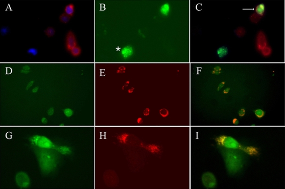
Figure 10. Membrane-anchored FRP-3 binds EGF at the cell surface.(A–C) Antibody staining for HA (in red, A,C) in CHO cells transfected with pCS2FRP3-HACD4 shows FRP-3 expression on the cell surface. Upon administration of EGFAlexa488, EGF expression (green) is detectable only on whole intact CHO cell expressing FRP-3, but not on control cells (C). Star in panel B shows non-specific EGF binding on necrotic cell. Double labelling for HA and GFP shows co-localization of FRP-3 and EGF at the cell surface (arrow in C). (D–I) Anti-EGF antibody staining (in red) on FRP-3GFP CHO cells transfected with pTWEENFRP3-HACD4. (D–F) Upon DTSSP cross-linking all CHO cells that express FRP-3CD4 (green staining in D) bind EGF at the membrane surface (red staining in E and merged colour in F). (G–I) 40× magnification of a single CHO cell that expressed FRP-3GFP (in green) and bind EGF (in red). Double labelling for GFP and anti-HA in panel I shows the two proteins interaction occurs at the membrane surface.
Image published in: Scardigli R et al. (2008)
Scardigli et al. Creative Commons Attribution license
Permanent Image Page
Printer Friendly View
XB-IMG-123014