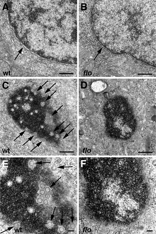
Figure 5. Nuclear ultrastructure in flo mutants.Transmission electron micrographs of nuclei from representative 5 dpf wild type and flo intestinal epithelial cells. (A,B) Intact nuclear envelope in wild type (A) and flo (B). (C–F) Tangential sections through the nuclear envelope showing abundant nuclear pores (arrows) in the wild type larva (C,E) but few if any well defined pores in the flo larva (D,F). (E) and (F) are higher magnification views of (C) and (D), respectively.
Image published in: Davuluri G et al. (2008)
Davuluri et al. Creative Commons Attribution license
Permanent Image Page
Printer Friendly View
XB-IMG-123382