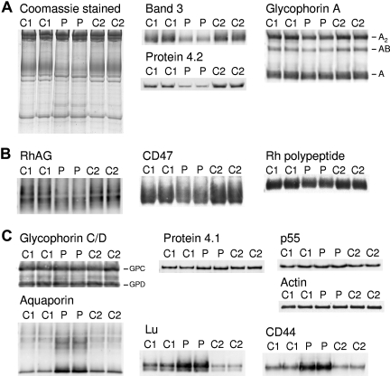
Figure 2. Coomassie and immunostaining of erythrocyte membrane proteins. Erythrocyte membranes were separated on 10% Laemmli gels and immunoblotted using antibodies as shown. Loading C1, C2, controls 1 and 2, respectively. P indicates proband. (A) Proteins of the band 3 complex: immunoblotting used the monoclonal antibody BRIC170 (N-terminal band 3) and antipeptide antibodies against C-terminal of protein 4.2 and GPA. (B) Proteins of the Rh complex: immunoblotting used antipeptide antibodies against C-terminal of RhAG, Rh polypeptides, and CD47. (C) Proteins of the glycophorin C (GPC) complex: immunoblotting used antipeptide antibodies against C-terminal of GPC and GPD, protein 4.1, and p55. Other proteins: immunoblotting used monoclonal antibodies anti–β-actin (Abcam), BRIC235 (CD44), and BRIC221 (Lu), and an antipeptide antibody against C-terminal aquaporin (AQP1). All antibodies used as described in “Erythrocyte membrane protein analysis.”
Image published in: Toye AM et al. (2008)
© 2008 by The American Society of Hematology. Creative Commons Attribution-NonCommercial license
Permanent Image Page
Printer Friendly View
XB-IMG-123506