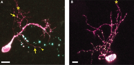
Figure 1. Distribution of CPEB- and delCPEB-containing RNP granules in optic tectal neurons. Optic tectal neurons labeled with cytosolic YFP (magenta) and CPEB–CFP (A) or delCPEB–CFP (B). CFP-tagged RNP granules are green puncta that when overlapping with magenta, appear white. Labeled RNP granules are distributed throughout the dendritic arbor of all cells imaged, whereas axons in 25% of the cells contain RNP granules. Examples of puncta in the locally branching axon (axons indicated with asterisks): arrows point to puncta within the axon arbor and arrowhead points to punctum at an axon tip. Scale bars = 10 μm.
Image published in: Bestman JE and Cline HT (2009)
Image downloaded from an Open Access article in PubMed Central. Copyright © 2009 Bestman and Cline.
Permanent Image Page
Printer Friendly View
XB-IMG-124235