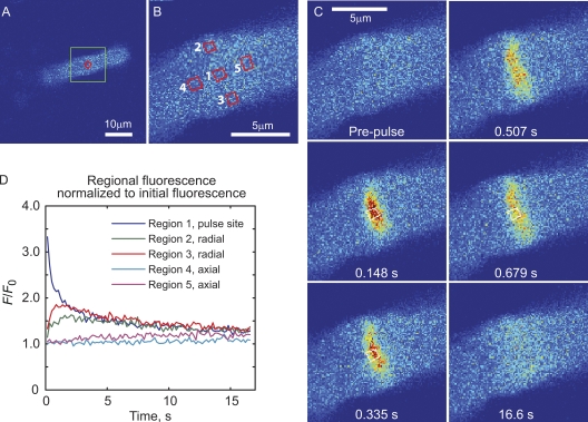
Figure 7. Equilibration of PAGFP in the OS compartment is highly anisotropic. (A) x–y image of the region of retinal slice showing the central z level of the cell at which the experiment was performed. The region of the OS that was rapidly scanned before and after a 100-µs photoconversion pulse (indicated by the red symbol) is delineated by the green box. (B) Prepulse scan of the region showing subregions where time courses of fluorescence change were recorded (red boxes). (C) Selected time course images showing the rapid radial and slower axial equilibration. (D) Time courses of fluorescence changes recorded from the regions shown in B. Radial positions 2 and 3 changed approximately in parallel and merge with the fluorescence time course from region 1, the site of photoconversion, within ∼2 s. The fluorescence at axial positions 4 and 5 required much longer, >15 s, to merge with the fluorescence of region 1, demonstrating the high degree of anisotropy in PAGFP diffusion in the OS. See Video 3.
Image published in: Calvert PD et al. (2010)
© 2010 Calvert et al. Creative Commons Attribution-NonCommercial-ShareAlike license
Permanent Image Page
Printer Friendly View
XB-IMG-124735