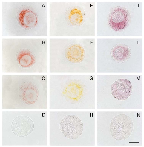
Figure 2. Lysosomal damage promoted by YTX in IPLB-LdFB cell lines evidenced by neutral red (A–D), acridine orange (E–H) and acid phosphatase activity (I–N) methods. A, E, I) IPLB-LdFB control cells; B, F, L) IPLB-LdFB cells after 8 h incubation with 100 nM YTX; C, G, M) IPLB-LdFB cells after 12 h incubation with 100 nM YTX; D, H, N) IPLB-LdFB cells after 24 h incubation with 100 nM YTX. Bar = 10 μm. (Reprinted with permission from [30]).
Image published in: Franchini A et al. (2010)
© 2010 by the authors; licensee Molecular Diversity Preservation International, Basel, Switzerland. Creative Commons Attribution license
Permanent Image Page
Printer Friendly View
XB-IMG-124827