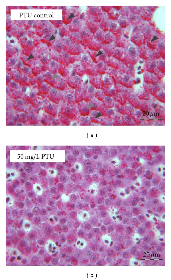
Figure 12. Representative histopathological alterations in hepatocytes of control (a) and PTU-exposed zebrafish (b). The well-visible glycogen deposits in the control ((a): ▲) are depleted in the higher concentration groups. Sections of 2 μm thickness stained with periodic acid-Schiff (PAS) and Mayer's hematoxylin.
Image published in: Schmidt F and Braunbeck T (2011)
Image downloaded from an Open Access article in PubMed Central. Copyright © 2011 F. Schmidt and T. Braunbeck.
Permanent Image Page
Printer Friendly View
XB-IMG-126328