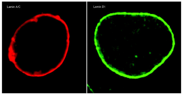
Figure 4. Nuclear lamins: localization at the nuclear periphery and within the nucleoplasm. Immunofluorescence staining of lamin A/C (red) and lamin B1 (green) in U2OS human osteosarcoma cell and MEF cell nuclei, respectively.
Image published in: Dittmer TA and Misteli T (2011)
Image downloaded from an Open Access article in PubMed Central. Copyright ©2011 BioMed Central Ltd.
Permanent Image Page
Printer Friendly View
XB-IMG-126633