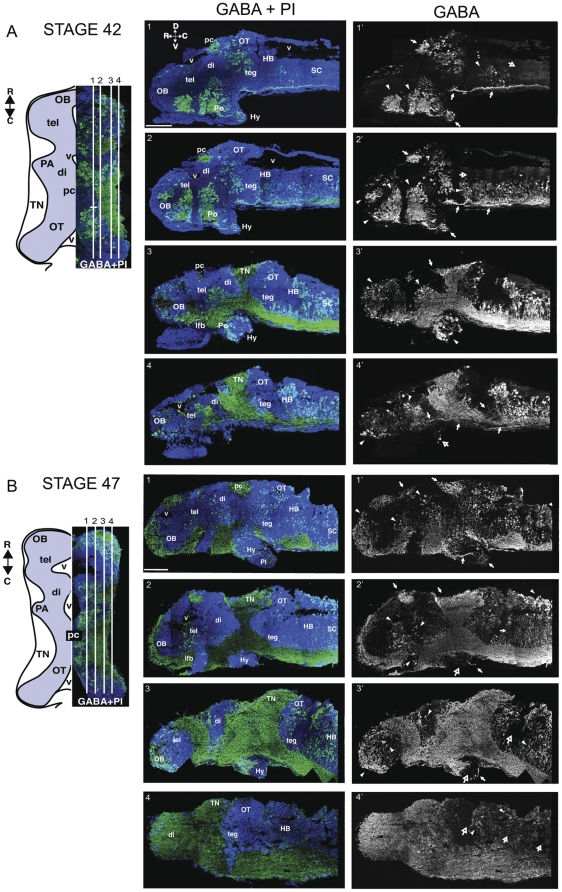
Figure 2. Distribution of GABA immunoreactivity in stage 42 and stage 47 tadpole CNS.A. Stage 42: Schematic (left) indicating relative positions of montaged sagittal sections of the tadpole brain. Blue is the cell body area; white is the neuropil area. Sections (1â4) show GABA immunostaining (green) counterstained with the nuclear label, propidium iodide (PI, blue). GABA staining alone is presented in the right panels (1â²â4â²). In panels 1â²â4â² arrowheads indicate GABA containing somata, filled arrows are GABA-positive axon tracts, and open arrows denote GABA-sparse zones. B. Sagittal series through a stage 47 tadpole brain. The pattern of GABA-immunoreactivity in the brain is similar to stage 42 except for a dispersion of the dense clusters of GABA immunoreactive cells seen in younger brains and the vast expansion of the labeled cell body regions, neuropil and axon tracts in the older tadpoles. Scale bars, 250 µm. See text for details.
Image published in: Miraucourt LS et al. (2012)
Miraucourt et al. Creative Commons Attribution license
Permanent Image Page
Printer Friendly View
XB-IMG-126775