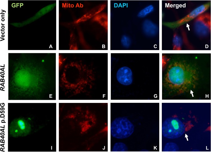
Figure 5. p.D59G disrupts normal intracellular protein localisation. Cells transiently transfected with N-terminal green fluorescent protein (GFP)-tagged vector constructs (vector control, A-D; RAB40AL, E-H; and RAB40AL p.D59G, I-L) and stained with anti-mitochondrial COXIV antibody (Mito Ab) and DAPI are shown. GFP–RAB40AL localises to the mitochondria and throughout the cytoplasm (panel H), while this localisation is disrupted with GFP–RAB40AL–p.D59G (panel L). GFP–RAB40AL–p.D59G appears to be accumulated or clustered within the nucleus, nucleolus and/or perinuclear region (panels I and L). Representative transfected cells are shown (arrows).
Image published in: Bedoyan JK et al. (2012)
© 2012, Published by the BMJ Publishing Group Limited. Creative Commons Attribution-NonCommercial license
Permanent Image Page
Printer Friendly View
XB-IMG-127171