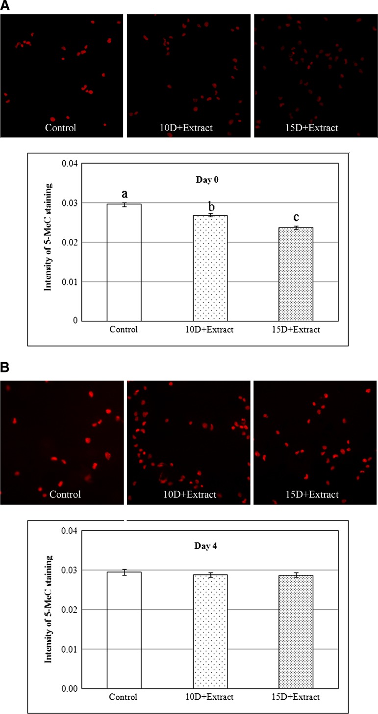
FIG. 2. Quantitative analysis of the DNA methylation status detected by 5MeC immunostaining in the control and Xenopus egg extract-treated cells (10D+extract: 10 μg/mL digitonin permeabilization of cells and Xenopus egg extract treatment; 15D+extract: 15 μg/mL digitonin permeabilization and Xenopus egg extract treatment) at days 0 (A) and 4 (B). The level of methylation is reported in arbitrary units and was detected using Texas Red-labeled antibodies (second) bound to primary antibodies specific to 5MeC. a, b, and c: Intensity of 5Mec staining in columns with different letters differ (a, b: p<0.05; a, c and b, c: p<0.01). 5MeC, 5-methylcytosine.
Image published in: Yang X et al. (2012)
Image downloaded from an Open Access article in PubMed Central. Copyright 2012, Mary Ann Liebert, Inc.
Permanent Image Page
Printer Friendly View
XB-IMG-128273