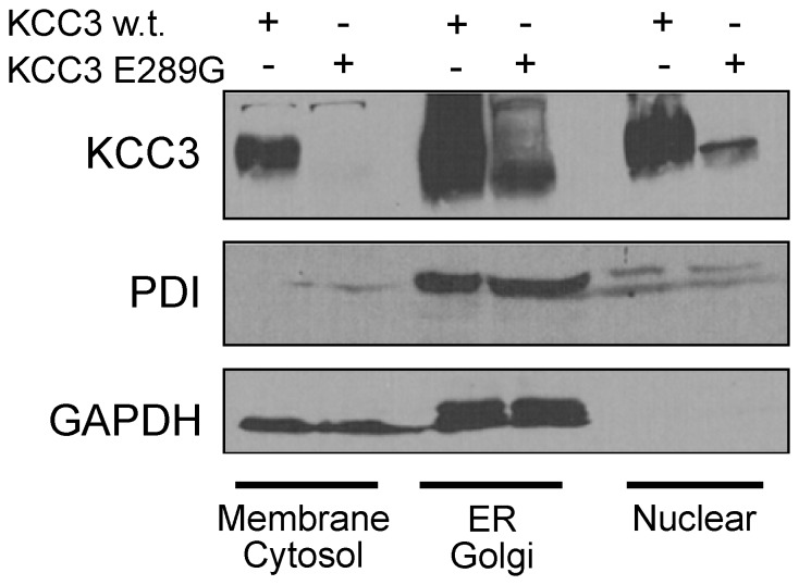
Figure 9. Sub-cellular localization of KCC3 and KCC3-E289G in HEK 293FT cells.HEK 293FT cells were transfected with wild-type KCC3 or KCC3-E289G mutant. Two days post-transfection, the cells were treated with digitonin to extract proteins from cholesterol-rich (plasma) membranes (membrane/cytosol fraction), followed by NP40 treatment to isolate proteins from ER/Golgi fraction, followed by deoxycholate+SDS detergents to isolate proteins from nuclear fraction. Western blots were probed with rabbit polyclonal anti-KCC3 and mouse monoclonal anti-PDI and anti-GAPDH antibodies. Experiment was reproduced at least 5 times with similar data.
Image published in: Ding J et al. (2013)
Image reproduced on Xenbase with permission of the publisher and the copyright holder. Creative Commons Attribution license
Permanent Image Page
Printer Friendly View
XB-IMG-128535