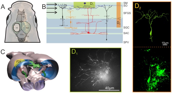
Figure 2. Morphology of neurons in the optic tectum of adult zebrafish.A. Image of the head of an adult zebrafish following exposure of the left tectum opticum. A schematic drawing of the brain is superimposed on the image of the head of a zebrafish. The region exposed for imaging is marked as the region of interest (ROI). B. Schematic transversal section through the tectum opticum (redrawn after [30]) showing the input layer and the overall cytoarchitecture of some prominent neurons. Areas highlighted in colour indicate where photomicrographs of labelled cells shown in D1âD2 are located. C. Schematic three-dimensional view on the tectum. The ROI is highlighted in yellow, while the tectal areas are indicated in blue (BioVis3D). D1âD2. Photomicrographs of stained cells. D1 shows an example of a top view using the in vivo imaging set-up (widefield fluorescence). D2 is an example of a neuron recovered after the in vivo experiments following routine histological methods. Here a confocal stack of the vibratome sectioned tectum was used to reconstruct the 3D properties of this neuron (BioVis3D). Abbreviations: SM Stratum moleculare; SO Stratum opticum; SFGS Stratum griseum et album superficial; SGC Stratum griseum central; SAC Stratum album central; SPV Stratum periventriculare; OB Olfactory bulb; Tel Telencephalon; TeO Tectum opticum; CCe Corpus Cerebellare; EG Eminentia granularis; MO Medulla oblongata.
Image published in: Kassing V et al. (2013)
Image reproduced on Xenbase with permission of the publisher and the copyright holder. Creative Commons Attribution license
Permanent Image Page
Printer Friendly View
XB-IMG-128639