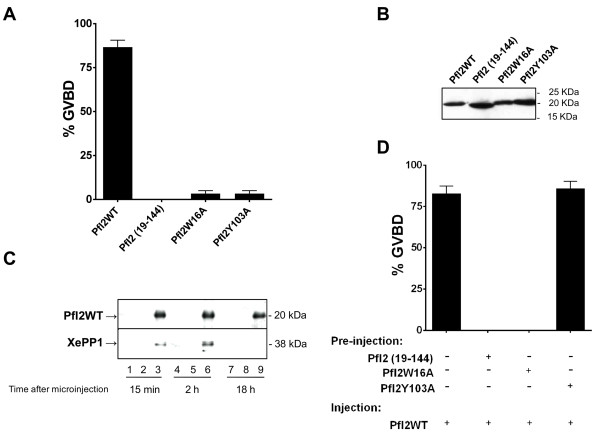
Figure 6. Initiation of G2/M transition in Xenopus oocytes by PfI2. Appearance of GVBD was monitored for 15 h after injection, and values are presented as percentages. Each experiment was performed using a set of 20 oocytes and was repeated at least three times. A. Percentage of GVBD induced with 100 ng of PfI2WT, PfI2(19–144), PfI2W16A or PfI2Y103A recombinant proteins. B. Immunoblot analysis of extracts prepared from injected oocytes revealed with mAb anti-His showing the presence of each recombinant protein after microinjection C. Interaction of PfI2 with Xenopus PP1. Immunoblot analysis using specific anti-XePP1 antibodies of naïve (lanes 1,4,7) or PfI2WT injected oocytes extracts after co-immunoprecipitation with anti-His mAb (lanes 3,6,9) (or anti-rabbit used as control (lanes 2,5,8)) revealed the presence of XePP1 in the complex (lanes 3,6). D. Percentage of GVBD induced by the pre-injection of PfI2(19–144), PfI2W16A or PfI2Y103A (100 ng) 2 hours before PfI2WT injection (100 ng). PfI2WT injection was used as a positive control.
Image published in: Fréville A et al. (2013)
Copyright ©2013 Frèville et al. Creative Commons Attribution license
Permanent Image Page
Printer Friendly View
XB-IMG-128999