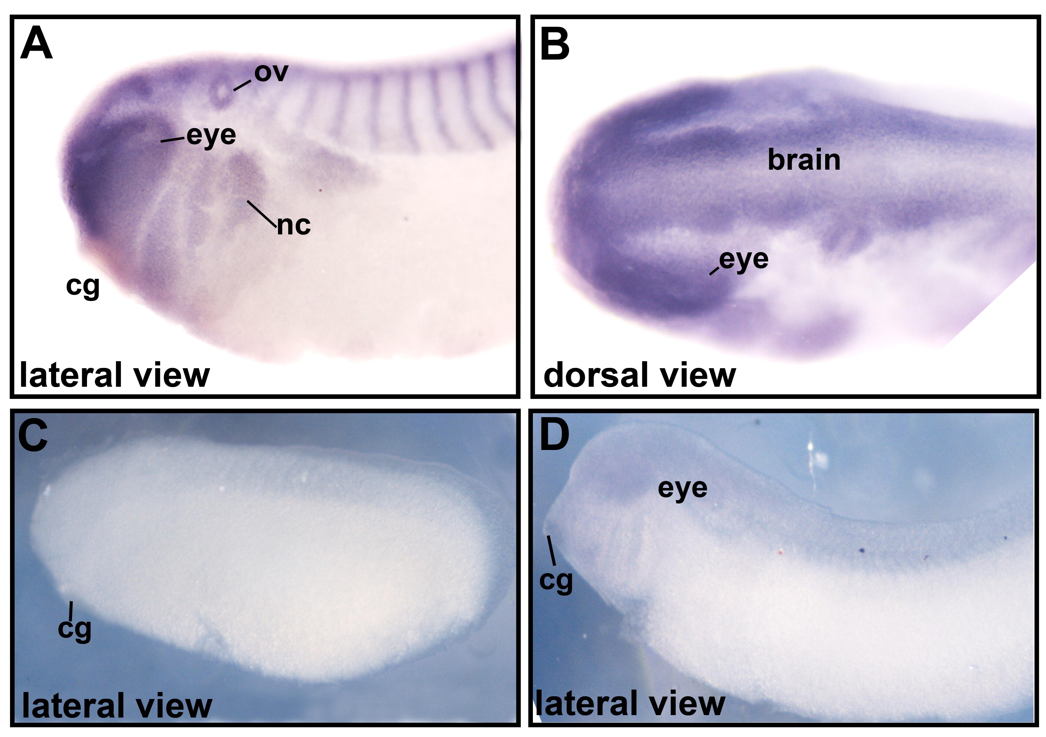
Supplementary Figure 2. A,B) Xenopus laevis in situ hybridization using a probe generated from the 3âUTR of the Xenopus tropicalis rai1 sequence. The region shares a 66% nucleotide similarity with the same region of the X. laevis rai1 gene and showed a highly similar staining pattern in X. laevis embryos. The probe sequence did not match any other sequence in either Xenopus species with identity of greater than 20% or with a stretch of nucleotides longer than 50. C,D) The sense or negative control probe show no significant staining pattern. A blue background was necessary to show contrast with the starkly white embryos.
Image published in: Tahir R et al. (2014)
Copyright © 2014. Image reproduced with permission of the Publisher.
| Gene | Synonyms | Species | Stage(s) | Tissue |
|---|---|---|---|---|
| rai1.S | smcr, sms | X. laevis | Throughout NF stage 28 | brain otic vesicle migratory neural crest cell anterior branchial crest posterior branchial crest cranial neural crest mandibular crest hyoid crest midbrain-hindbrain boundary olfactory placode pronephric mesenchyme intersomitic region neural tube forebrain midbrain hindbrain |
Image source: Published
Permanent Image Page
Printer Friendly View
XB-IMG-130154