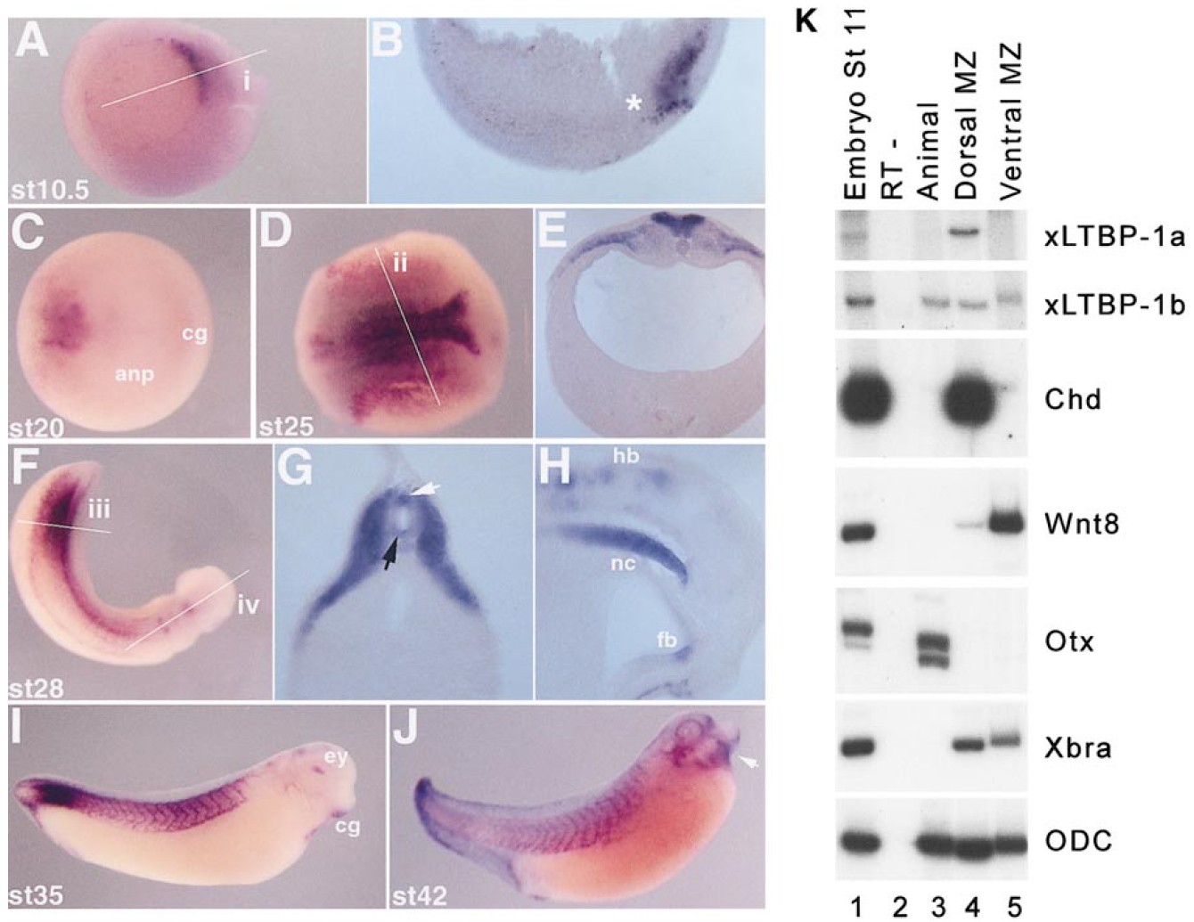
FIG. 3. In situ hybridization of Xenopus LTBP-1. (AâJ) In situ hybridization. (A): Stage 10.5 (gastrula). Expression of xLTBP-1 is detected within the organizer region. (B) A section (depicted by the line âiâ in A) shows expression is restricted to the dorsal mesoderm of the organizer (*). (C) Stage 20. Expression of xLTBP-1 in the spinal cord. No expression is seen in the anterior neural plate at this stage. Anterior/ventral is to the right. (D) Stage 25. Transcripts can be detected throughout the spinal cord region but still not in anterior domains. (E) A section through the embryo at the level of the spinal cord (indicated by the line âiiâ in D); xLTBP-1 RNA expression is seen throughout the neural tube but not in the notochord at this stage. (F) Expression is also detected in the lateral plate mesoderm, Stage 28. Expression is seen in the tail, throughout the spinal cord and in the hindbrain. (G) Transverse section through the spinal cord (âiiiâ in F); xLTBP-1 RNA is expressed in the mesoderm lateral to the neural tube, in the notochord and in both the roof (white arrow) and floor plates (black arrow). (H) Midsagital section through the head (line âivâ in F). xLTBP-1 RNA is also expressed in anterior regions, in the hindbrain, and in the ventral forebrain. Note the expression within the notochord. (I) Stage 35. xLTBP-1 RNA is highly expressed in the muscle blocks surrounding the spinal cord. It is also expressed in the rhombomeres of the hindbrain, the eye, and the cement gland. High expression is present in the tailbud cordoneural hinge. (J) Stage 42. Additional xLTBP-1 RNA expression is observed in more anterior regions, including the heart (white arrow), the branchial arches, and around the eye. anp, anterior neural plate; cg, cement gland; ey, eye; fb, forebrain; hb, hindbrain; he, heart; nc, notochord. Locations of the sections are indicated by dashed lines. (K) xLTBP-1 RNA expression in dissected gastrula embryos: Dorsal and ventral marginal zones and animal cap explants were dissected from stage 11 gastrula embryos and analyzed by RT-PCR. xLTBP-1 was amplified by using the same primers as in Fig. 2 but for 30 cycles. The following controls are included as markers for the dissected fragments: Chordin (chd) for dorsal mesoderm, Wnt8 for ventral mesoderm, Otx for animal cap, Brachyury (xBra) as a pan mesodermal marker, and ODC as the loading control.
Image published in: Altmann CR et al. (2002)
Copyright © 2002. Image reproduced with permission of the Publisher, Elsevier B. V.
| Gene | Synonyms | Species | Stage(s) | Tissue |
|---|---|---|---|---|
| ltbp1.S | LTGF-1, TGFbBP1 | X. laevis | Throughout NF stage 10.5 | upper blastopore lip involuted dorsal mesoderm |
| ltbp1.S | LTGF-1, TGFbBP1 | X. laevis | Throughout NF stage 20 | neural tube |
| ltbp1.S | LTGF-1, TGFbBP1 | X. laevis | Throughout NF stage 25 | lateral plate mesoderm neural tube |
| ltbp1.S | LTGF-1, TGFbBP1 | X. laevis | Throughout NF stage 28 | forebrain hindbrain notochord paraxial mesoderm presomitic mesoderm somite |
| ltbp1.S | LTGF-1, TGFbBP1 | X. laevis | Throughout NF stage 35 and 36 | cement gland muscle eye presomitic mesoderm brain heart somite |
| ltbp1.S | LTGF-1, TGFbBP1 | X. laevis | Throughout NF stage 42 | somite pharynx cranial nerve vagus nerve ophthalmic vein mouth primordium otic vesicle |
Image source: Published
Permanent Image Page
Printer Friendly View
XB-IMG-130910