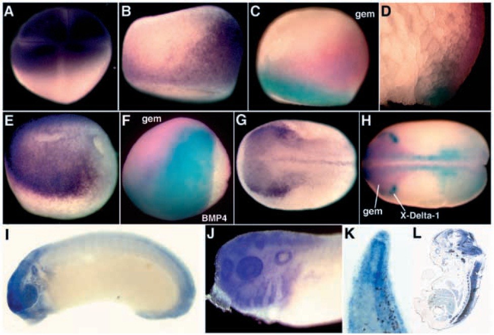
Fig. 4. Spatial distribution of geminin transcripts in the early embryo identified by in situ hybridization. (A,B,E,G,I-K) Geminin staining is purple in singly stained embryos. (C,D,F,H) Geminin expression is pink in doubly stained embryos. The expression patterns stained in aqua are brachyury (C,D), BMP4 (F), or X-Delta-1 (H). (L) Geminin expression detected in a section of an e15 mouse embryo. Dorsal is oriented to the right (B-D), top (E,F) or facing out (G,H). Xenopus embryos are stages 4 (A), 10 (B), 10.25 (C, D), 12 (F), 12.5 (E), 13.5 (G), 20 (H), 28 (I) and 38 (J,K).
Image published in: Kroll KL et al. (1998)
Copyright © 1998. Image reproduced with permission of the Publisher and the copyright holder. This is an Open Access article distributed under the terms of the Creative Commons Attribution License.
Permanent Image Page
Printer Friendly View
XB-IMG-131466