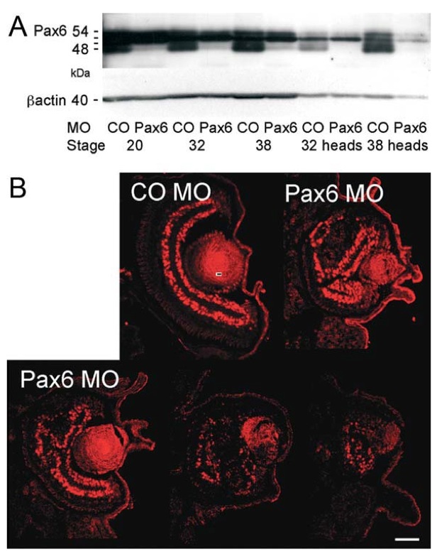
Figure 1 Reduction of Pax6 protein by morpholino treat- ment. A: Western blot using polyclonal Pax6 antibodies on proteins from whole embryos carrying control morpholino (MO CO) or Pax6 antisense-morpholino (MO Pax6), or from isolated heads at developmental Stages 20, 32, and 38. Immunoreactive bands migrate between Mr 48 and 54 kD. Aliquots of the same extracts were analyzed for bactin (lower panel). B: Immunofluorescent staining for Pax6 on eyes from control (CO MO) and Pax6-inhibited (Pax6 MO) Stage 42 tadpoles. Small-eyed phenotypes show retinal folding (top right, bottom left) and duplications (bottom middle). Rudimentary eye anlagen contain a reduced num- ber of Pax6 expressing cells (bottom right). Bar, 50 um. [Color figure can be viewed in the online issue, which is available at wileyonlinelibrary.com.]
Image published in: Rungger-Brändle E et al. (2010)
Copyright © 2010. Image reproduced with permission of the Publisher, John Wiley & Sons.
| Gene | Synonyms | Species | Stage(s) | Tissue |
|---|---|---|---|---|
| pax6.L | an2, mgda, pax-6, pax6-a, pax6-b, wagr, XLPAX6, xpax6 | X. laevis | Throughout NF stage 42 | retina eye lens retinal neural layer |
Image source: Published
Permanent Image Page
Printer Friendly View
XB-IMG-133613